
Figure 3.
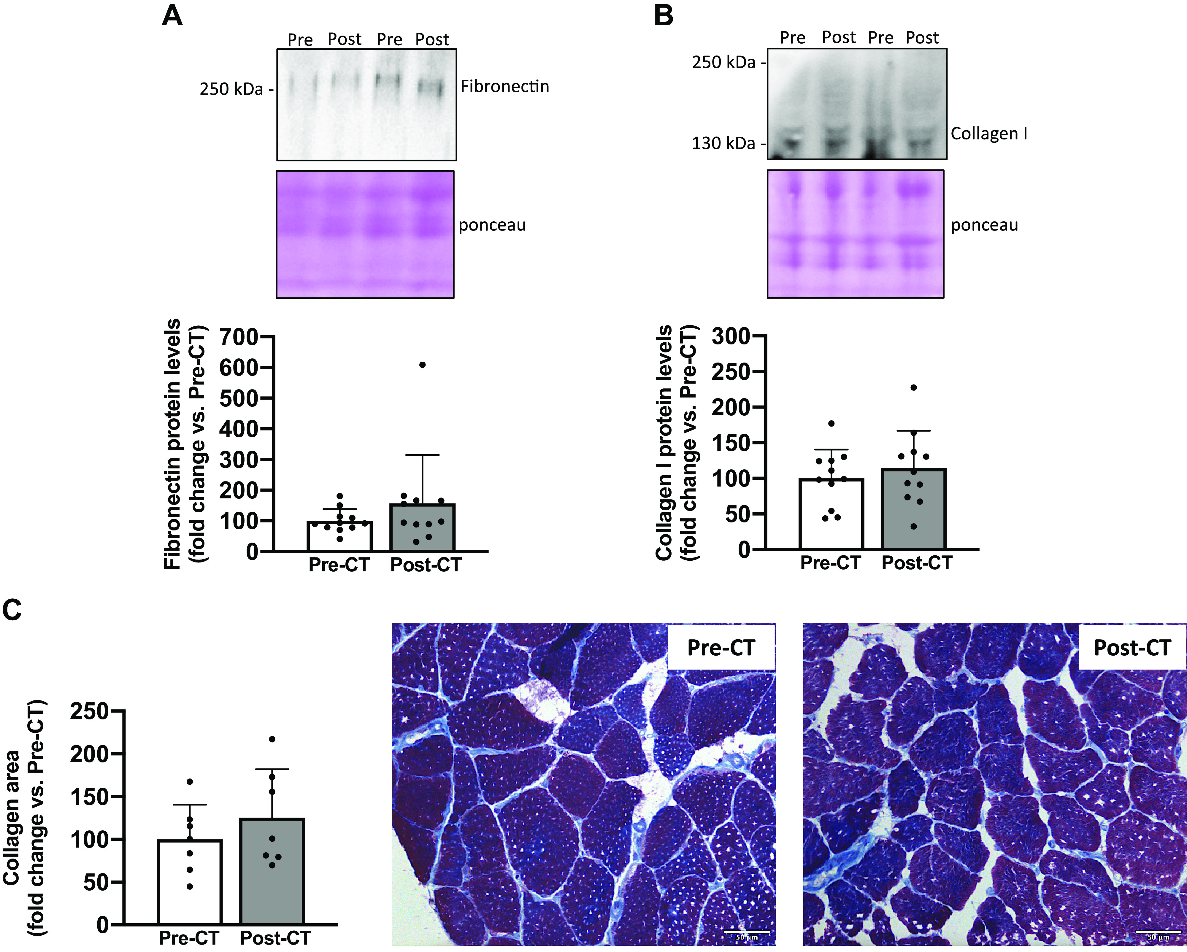
Changes in markers of fibrosis. Fibronectin (A) and collagen I (B) protein levels from vastus lateralis muscle biopsies taken before (Pre) and after (Post) chemotherapy (CT) (n = 11). For each Western blot, two representative subjects are displayed. Modified Masson’s trichrome staining from transverse cross sections of vastus lateralis muscle biopsies pre- and post-CT and representative images of muscle transversal sections with collagen stained in blue (C). Biopsy samples from four patients were of poor quality and were excluded from histological analysis (n = 7). All values are expressed as the means ± SD. *P < 0.05.