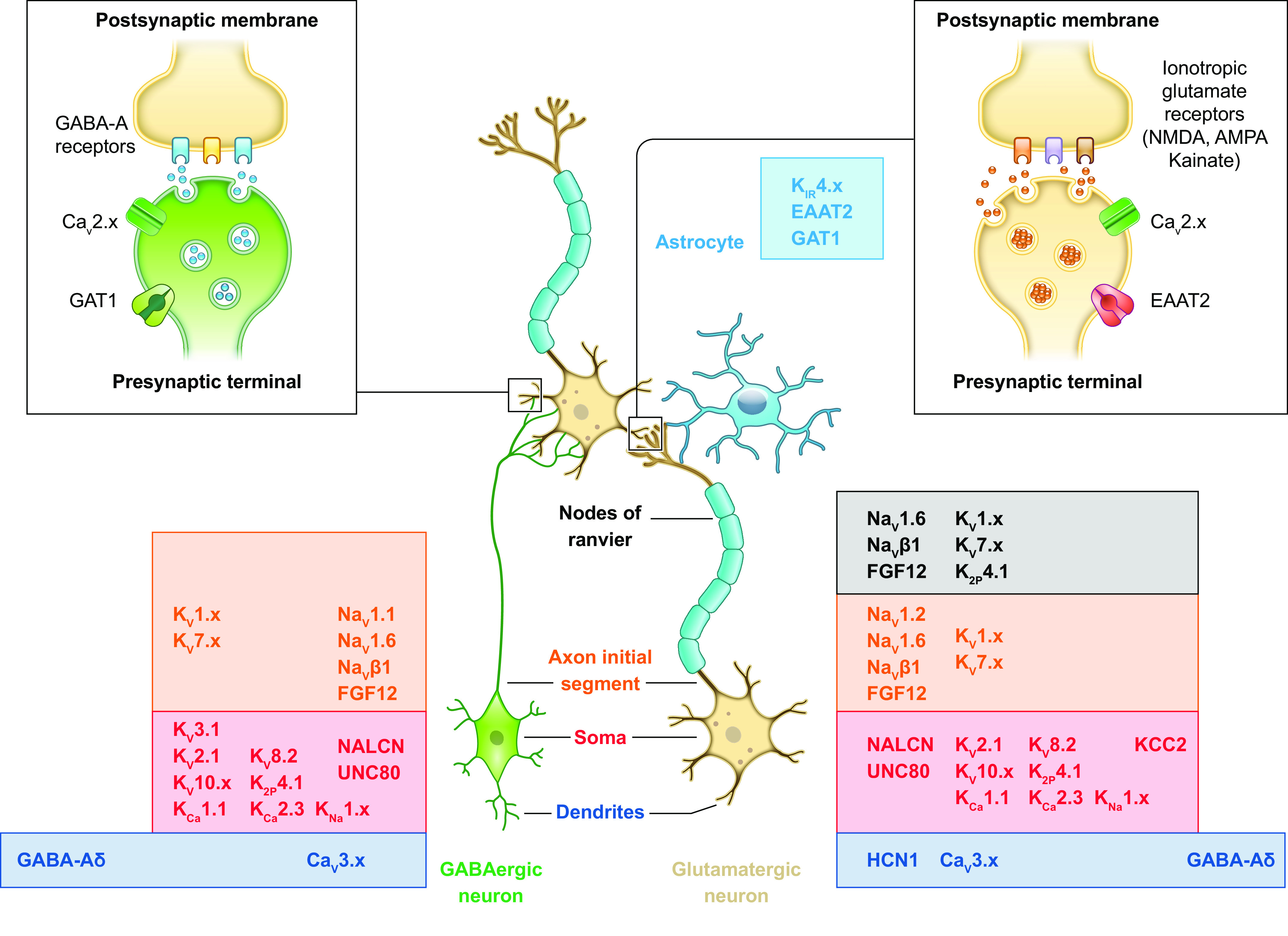
FIGURE 5.

Simplified diagram of a cortical microcircuit with interconnected glutamatergic and GABAergic neurons, an astrocyte, and cellular/subcellular distribution of ion channels and transporters. A cortical neuronal microcircuit is illustrated as a presynaptic GABAergic neuron (green) and a presynaptic myelinated glutamatergic neuron (ocher) that form synaptic connections on the dendrites of a myelinated glutamatergic neuron (ocher). Glial cells are displayed as an astrocyte (light blue) in proximity of the glutamatergic synapses and as the myelin sheets around the axons of the glutamatergic neurons formed by oligodendrocytes (violet; the soma is not displayed), allowing saltatory conduction at the nodes of Ranvier. The insets at top show in more detail a GABAergic (left) and a glutamatergic (right) synapse. The ion channels and transporters targeted by DEE mutations are indicated with their protein name (see text for details) and their known cellular/subcellular distribution, according to the neuronal subcompartments (dendrites, soma, axon initial segment, nodes of Ranvier of the myelinated axon, presynaptic terminal, and postsynaptic membrane).
