FIGURE 7.
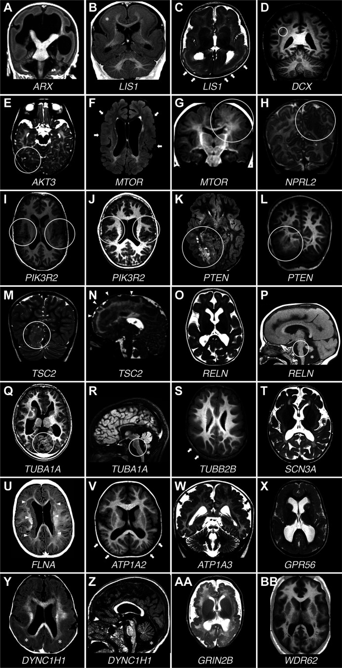
Brain MRI of patients with different malformations of cortical development. A: T1-weighted (T1W) coronal section. Lissencephaly in a boy with ARX mutation. The ventricles are severely dilated, the corpus callosum is absent, and the basal ganglia are severely hypoplastic. B and C: coronal T1W and axial T2-weighted (T2W) sections of a brain with posterior > anterior pachygyria and increased cortical thickness. Boy with LIS1 mutation. White asterisk in B indicates the point of more severe cortical thickening. White arrows in C point to areas of more severely smooth and thick cortex. D: T1W coronal section. Diffuse subcortical band heterotopia in a girl with DCX mutation. White circle surrounds the subcortical laminar heterotopia, which forms an almost continuous band beneath the cortex, separated from it by white matter. E: axial T2W section. Right occipital cortical dysplasia (surrounded by a white circle) in a girl with a very low-level mosaic mutation in AKT3 (0.67% in brain, not detectable in blood). F and G: axial fluid-attenuated inversion recovery (FLAIR) and coronal T1W sections in 2 patients carrying mosaic mutations in the MTOR gene with different % of mosaicism (F: p.Thr1977Ile, 20% of mosaicism in blood; G: p.Ser2215Phe, 5.5% of mosaicism in the surgically removed dysplastic brain tissue). In F, the patient has megalencephaly with large ventricles and multiple areas of abnormal cortex alternating infoldings with smooth surface. This pattern is suggestive of polymicrogyria (white arrows). In G, white circle highlights an area of cortical dysplasia with increased volume of the brain parenchyma, blurring of the gray-white matter junction, and irregular cortical folding. H: T2W coronal section. Left parieto-temporal focal cortical dysplasia in a girl with NPRL2 mutation. Circle surrounds the parietal portion of the cortical abnormality. I and J: T1W axial sections in 2 patients carrying the p.Gly373Arg PIK3R2 gene mutation with different % of mosaicism (I: 13% of mosaicism in blood, 43% in saliva; J: 10% of mosaicism in blood, 29% in saliva). Both patients have bilateral perisylvian polymicrogyria (white circles). K and L: axial FLAIR and coronal T1W sections showing right posterior quadrantic dysplasia (white circle) in a boy with a constitutional PTEN mutation. M and N: T2W coronal and sagittal sections in 2 patients with constitutional TSC2 mutations (M: p.Thr1623Ile; N: p.Pro1202His) showing right posterior quadrantic dysplasia caused by a large cortical tuber (M, white circle) and an extensive dysplastic area involving most of the right frontal lobe (N, white arrowheads). O and P: T2W axial and T1W sagittal sections. Lissencephaly with normally thick cortex and cerebellar hypoplasia (P, asterisk) in a girl with RELN mutation. White circle surrounds a hypoplastic brain stem. Q and R: axial and sagittal T1W sections. Thickened cortex with simplified gyral pattern and cerebellar hypoplasia in a boy with TUBA1A mutation. Circles surround the hypoplastic cerebellum and brain stem. Asterisk indicates the area below a hypoplastic cerebellar vermis, and black arrow points to a hypoplastic corpus callosum lacking its most posterior part. S: T1W axial section. Diffusely simplified gyral pattern with prominent thickening and infolding of the sylvian fissures in a boy with TUBB2B mutation. Arrows point to an area of smooth cortex. T: T2W axial section. Severe dysgyria with simplified gyral pattern in a girl with SCN3A mutation. U: T1W axial section. Classical bilateral periventricular nodular heterotopia in a girl with FLNA mutation. Bilateral nodules of subependymal heterotopia (white arrowheads) are contiguous, extensively lining the ventricular walls. V: T1W axial section. Diffuse polymicrogyria, more prominent posteriorly (white arrows) in a boy with ATP1A2 mutation. W: T2W coronal section. Polymicrogyria with abnormal cortical infoldings and packed microgyria (black arrows), combined with abnormal sulcation in a boy with ATP1A3 mutation. X: T2W axial section. Bilateral frontoparietal cortical thickening and diffusely abnormal cortical pattern in a boy with biallelic GPR56 mutations. Y and Z: T1W axial and sagittal sections. Pachygyria and perisylvian polymicrogyria in a girl with DYNC1H1 mutation. Asterisks in Y are located where there is maximum cortical thickening, in the posterior cortex. Asterisk in Z is located beneath a hypoplastic cerebellar vermis. AA: T2W axial section. Diffuse polymicrogyria in a boy with a GRIN2B mutation. BB: T1W axial section. Diffuse abnormality of the cortical pattern with smooth cortex and areas of abnormal infolding, suggestive of polymicrogyria in a boy with biallelic WDR62 mutations.

