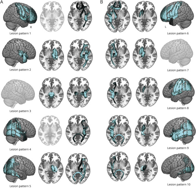
Figure 2. Individual Stroke Lesions Captured in Low Dimensions via Unsupervised Machine Learning Techniques.
Our automatically derived 10 unique lesion patterns represented mainly right- (A) and left-hemispheric (B) strokes. The patterns featured anatomically plausible collections of brain regions with varying emphases ranging from cortical-subcortical and anterior-medial-posterior regions. Of note, there were 5 clearly left-hemispheric lesion patterns and 4 right-hemispheric ones. Lesion pattern 3 primarily comprised the brainstem, yet was nonetheless assigned to right-hemispheric strokes because of right hemispherically more pronounced white matter tract contributions. Disregarding minor cortical and subcortical contributions, most right-hemispheric lesion patterns had a left-hemispheric pendant (i.e., lesion patterns 1 and 6, lesion patterns 2 and 6, lesion patterns 4 and 9, and lesion patterns 5 and 10). More specifically, lesion pattern 1 on the right and lesion pattern 6 on the left reflected infarcts involving mainly cortical territory, extending from the frontal pole to precentral and postcentral gyrus, including the insular and opercular cortex. The left-sided version of this pattern furthermore comprised subcortical nuclei, that is, the dorsal striatum. Lesion patterns 2 and 7 were mostly focused on subcortical nuclei, that is, thalamus and the basal ganglia as well the thalamic radiation and longitudinal and fronto-occipital WM tracts. Although the right-hemispheric pattern additionally gave weight to insular and opercular regions, there was no cortical contribution to the left-hemispheric one. Lesion pattern 3 was unique in predominantly representing the brainstem and predominantly right-hemispheric white matter tracts, with an emphasis on the corticospinal tract. Lesion pattern 8, on the other hand, featured left-hemispheric lesions in precentral and postcentral regions, insular, opercular, and parietal regions and did not have a direct analog among the right-hemispheric lesion patterns. In contrast, lesion patterns 4 and 9 reflected similarly configurated temporoparietal lesions in the right and left hemisphere, respectively. Their main difference related to the inclusion of subcortical nuclei in case of the left-hemispheric version. Finally, lesion patterns 5 and 10 depicted lesions with a focus on posterior circulation strokes, for example, including the visual cortex and fusiform gyrus. Individual patients' lesions were recreated by the weighted combination of lesion pattern. For example, a patient with a lesion in left subcortical brain regions would be characterized by high values for lesion pattern 7 but very low values for all other patterns. Renderings are shown in transparent in case of missing contributions from respective (sub)cortical regions.

