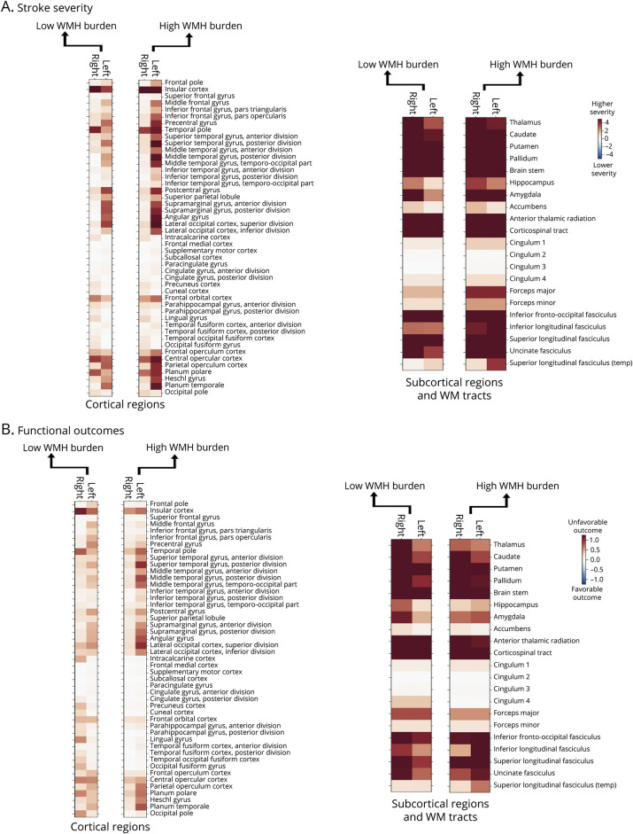
Figure 4. Relevance of Each Cortical and Subcortical Brain Region and White Matter Tract in Explaining NIHSS-Based Stroke Severity (A) and mRS-Defined Functional Outcomes (B) in the Derivation Cohort.
Darker red color translates to a more pronounced effect of a specific region, indicating a higher stroke severity or odds of unfavorable outcome, when lesioned. Results are grouped separately for cortical regions and subcortical regions/white matter tracts. For an eased comparison of results for patients with varying WMH burdens, results for patients with a lower WMH burden are illustrated on the left-hand side in direct proximity to results for patients, with a higher WMH burden on the right-hand side. Altogether, bilateral strokes affecting subcortical gray matter brain regions and white matter tracts explained higher stroke severity and higher odds of unfavorable outcomes in case of patients with both low and high WMH burden. In the case of stroke severity, left-lateralized cortical lesions had additionally relevant contributions that were more pronounced for patients with a high WMH burden (c.f., Figure 5). The effect of cortical lesions was altogether weaker in the case of functional outcomes. mRS = modified Rankin Scale; NIHSS = NIH Stroke Scale; WMH = white matter hyperintensity.

