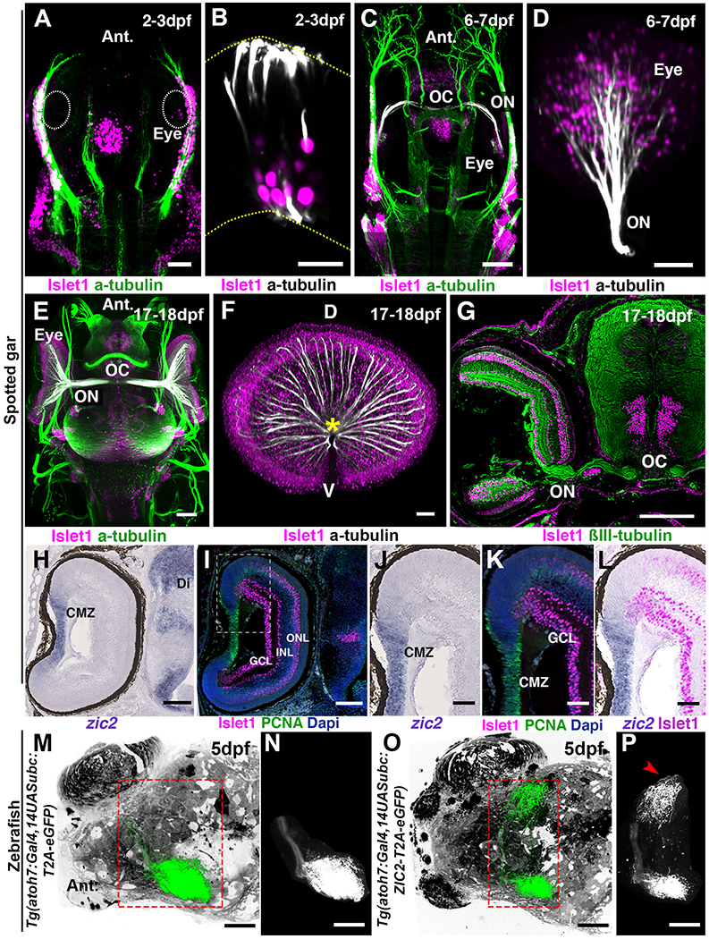
Fig. 4. Zic2 is not expressed by ganglion cells in spotted gar and zebrafish.

(A to F) Development of the visual system in the spotted gar. All images are 3D light-sheet fluorescence microscopy images of EyeDISCO-cleared spotted gar embryos labeled with Islet1 and acetylated Tubulin. (A and C and E) Top (dorsal) views of spotted gars at 2-3dpf (A), 6-7dpf (C), and 17-18dpf (E). (B and D and F) frontal views of whole spotted gar eyes at 2-3dpf (B), 6-7dpf (D), and 17-18dpf (F). The optic nerve (ON and asterisk) starts to form by 6-7dpf and is well developed by 17-18dpf. The optic chiasm (OC) is formed by 6-7dpf. (G) Coronal cryosection of spotted gar embryos at 17-18dpf labeled for ßIII-tubulin and Islet1. (H to K) cryosection from 17-18dpf spotted gar eyes hybridized with zic2 riboprobe (H and J) and labeled for proliferating cell nuclear antigen (PCNA) and Islet1 (I, K and L). (J to L) higher magnification of the ciliary margin zone (area framed in I). (M to P) 3D rendering of whole-brain viewed from the top of zebrafish injected with Tg(atoh7:Gal4,14UASubc:T2A-eGFP-pA) (M and N) or Tg(atoh7:Gal4,14UASubc:ZIC2-T2A-eGFP-pA (O and P). N and P show segmented ganglion cell projections. (P) A large ipsilateral projection (arrowhead) is seen in the Tg(atoh7:Gal4,14UASubc:ZIC2-T2A-eGFP-pA)-injected fish. Abbreviations: ON, Optic nerve; OC, Optic chiasm; GCL, Ganglion cell layer; INL, Inner nuclear layer; ONL, Outer nuclear layer. Scale bars are 50 μm in (A, D, F, J, K, L, M to P) and 15 μm in (B) and 80 μm in (C, H and I) and 150 μm in (E) and 200μm in (G).
