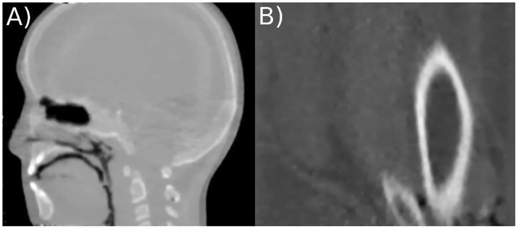
Figure 3:

Vertical slice of a HeCT of A) the CIRS pediatric head phantom model HN715 (CIRS, Norfolk, Virginia, USA) and B) the custom build animal tissue phantom reconstructed using the DROP-TVS iterative reconstruction algorithm with 256×256 pixel per slice and slice thickness of 1.25 mm. The phantom was 6 scanned using 90 projections consisting of ~4×10⁶ particles each.
