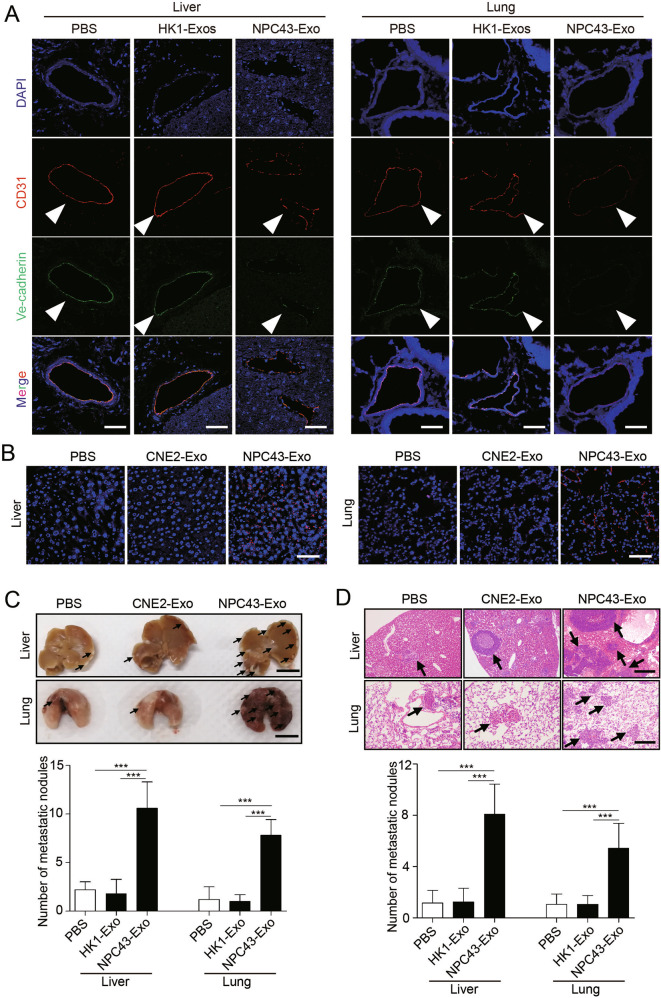
Fig. 2. Exosomes derived from EBV-positive NPC cells induce vascular permeability and promote metastasis in vivo.
A Collected lung and liver tissues were subjected to double-label immunofluorescence for VE-cadherin (green) and CD31 (red). Structures positive for CD31 are indicated by arrowheads. The scale bar represents 100 μm. B In vivo vascular permeability determined by the appearance of intravenously injected rhodamine-dextran in tissue (red) (n = 6). Representative images are shown. DAPI (blue): cell nuclei. The scale bar represents 100 μm. C Gross appearance of the liver and lung in the tail vein injection metastatic model and metastatic lesion quantitative analysis. Data are presented as the mean ± SD (n = 6 biologically independent animals). ***p < 0.001. D Representative hematoxylin and eosin (H&E) stained metastatic tumor nodules in the liver and lung, and quantitative analysis of metastatic nodules. Data are presented as the mean ± SD (n = 6 biologically independent animals). ***p < 0.001.

