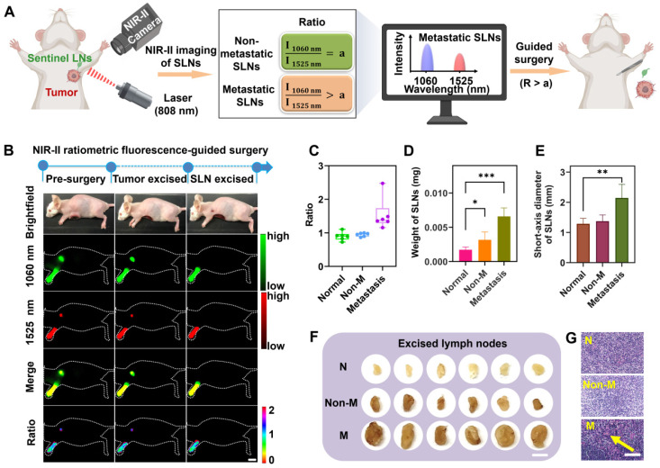
Figure 3.
Identification of the metastatic state of SLNs based on NIR-II ratiometric fluorescence. (A) Scheme of NIR-II ratiometric fluorescence probes for intraoperative detection and guided-surgery in orthotopic 4T1 breast cancer model. Images were created with BioRender.com. (B) NIR-II ratiometric fluorescence strategy for preoperative diagnosis and intraoperative navigation surgery in an orthotopic 4T1 breast cancer model. (C) Ratiometric signals (I1060 nm/I1525 nm) of SLNs in different groups, including (i) normal SLNs, (ii) non-metastatic SLNs, and (iii) metastatic SLNs (n = 6 for each group). (D) The weight of SLNs in all three groups (n = 6 for each group). Error bars mean ± SD, *P < 0.05, ***P < 0.001. (E) Short-axis diameter of SLNs in different groups (n = 6 for each group). Error bars mean ± SD, **P < 0.01. (F) Photograph of excised SLNs from three groups. N represents normal SLNs, Non-M represents non-metastatic SLNs, and M represents metastatic SLNs. The scale bar is 20 mm. (G) Representative H&E staining images of SLNs derived from different groups. Yellow arrows indicate foci occupied by tumor cells. The scale bar is 100 µm.

