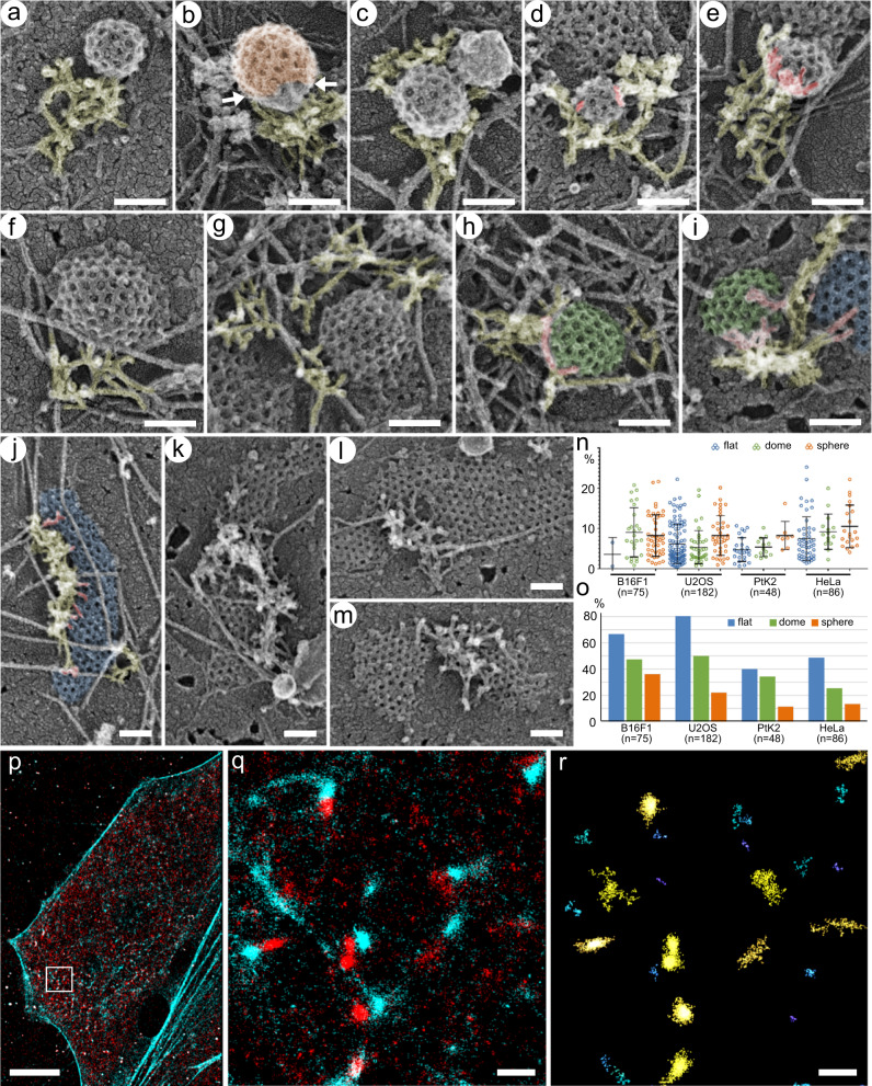
Fig. 2. Branched actin networks localize at the CCS perimeter with minimal overlaps with the clathrin lattice.
a–e Spherical CCSs in U2OS cells. Branched actin networks (yellow) can form a lateral comet tail (a, b, e), surround the CCS (d), or localize mainly underneath the CCS (c). b Branched actin network contacts an uncoated part (neck) of the CCS (clathrin coat is shown in orange). d, e Examples of actin filaments extending over the clathrin lattice (red); e shows a maximal observed level of actin-CCS overlap. f–i Dome-shaped CCSs in U2OS cells. Branched actin networks are laterally associated with CCSs (f, g) with occasional overhangs over the CCS periphery (h and i, red). i Branched actin network located in part at the interface between dome-shaped (green) and flat (blue) CCSs. j–m Flat CCSs in U2OS cells. Branched actin networks associate with the flat CCS (blue) margin (j), partially surround the CCS and separate it from the neighboring CCS (k), occupy a “bay” in the CCS (l), or localize between two neighboring CCSs (m), in all cases exhibiting minimal overhangs of actin filament ends over the CCS (red in j). n Percentage of the CCS area overlaid by branched actin filaments in 2D projection images within the actin-positive CCS subpopulations in the indicated cell types. Error bars, mean ± SD. o Fraction of the actin-positive CCSs with any amount of branched actin extending over their lattice in 2D projection images in the indicated cell types. p–r STORM imaging of intact fixed PtK2 cells stained with AlexaFluor488-phalloidin (cyan) and AlexaFluor405-647-labeled antibody against CHC (red). p Cell overview. q Zoomed region outlined by white box in p. r Region in q after Voronoi segmentation of clathrin localizations. Scale bars: 100 nm (a–m), 10 µm (p) and 500 nm (q, r).

