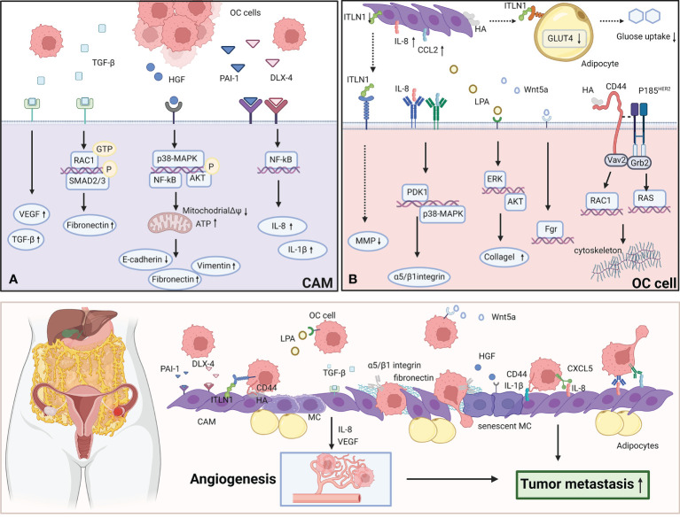
Figure 1.
CAMs interact with OC cells to promote the metastasis. (A) OC cells secret TGF-β, HGF, PAI-1, DLX-4 to effect CAMs via various signaling pathways. TGF-β activates RAC1/SMAD3 pathway via TGF-βRII to induce CAMs to upregulate fibronectin expression. HGF promotes the premature senescence of normal mesothelial cells by inducing mitochondrial oxidative stress via activating several signaling pathways including p38-MAPK, AKT and NF-κB. PAI-1 and DLX4 induce the expression of IL-8/CXCL5 and IL-1β/CD44 via activating NF-κB signaling. (B) CAMs overexpress ITLN1, IL-8, CCL2, LPA, Wnt5a and HA to effect OC cells by activating several signaling pathways. IL-8 induces the overexpression of PDK1 in OC cells via CXCR1.PDK1 upregulates the expression of α5 and β1 integrin to enhance the adhesion to fibronectin and mesothelial cells. CCL2 facilitates the trans-mesothelial migration and invasion of OC cells via activating p38-MAPK pathway through CCR2. Wnt5a boosts the metastasis of OC cells via activating its downstream effector Src family kinase Fgr. LPA activates ERK and Akt pathway to boost OC cells to adhere to collagen I. HA can bind to CD44v3-Vav2 complex on OC cells to activate Rac1 and Ras pathway signaling. The figure was created with BioRender.com.

