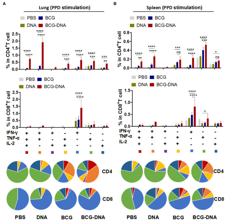
Figure 4.
Induction of Ag-specific polyfunctional T-cells in lungs and spleens of immunized mice. Six weeks after the primary immunization, the mice were sacrificed, and their lung and spleen cells were collected and treated with PPD (10 μg/ml) at 37°C for 15 h in the presence of GolgiStop. Ag-specific T cells secreting IFN-γ, TNF-α and IL-2 were distinguished by flow cytometry and classed into seven sub-populations based on the production of single or multiple cytokines. The percentages of the seven sub-populations as components of the total CD4+ or CD8+ T cells in lung (A) and spleen (B) and the pie chart analysis are shown. (n = 3 mice, two-way ANOVA, mean ± SEM). *p < 0.05, **p < 0.01, ***p < 0.001, ****p < 0.0001: a significant difference of treatment groups from the appropriate controls (PBS group). ns, no significant difference.

