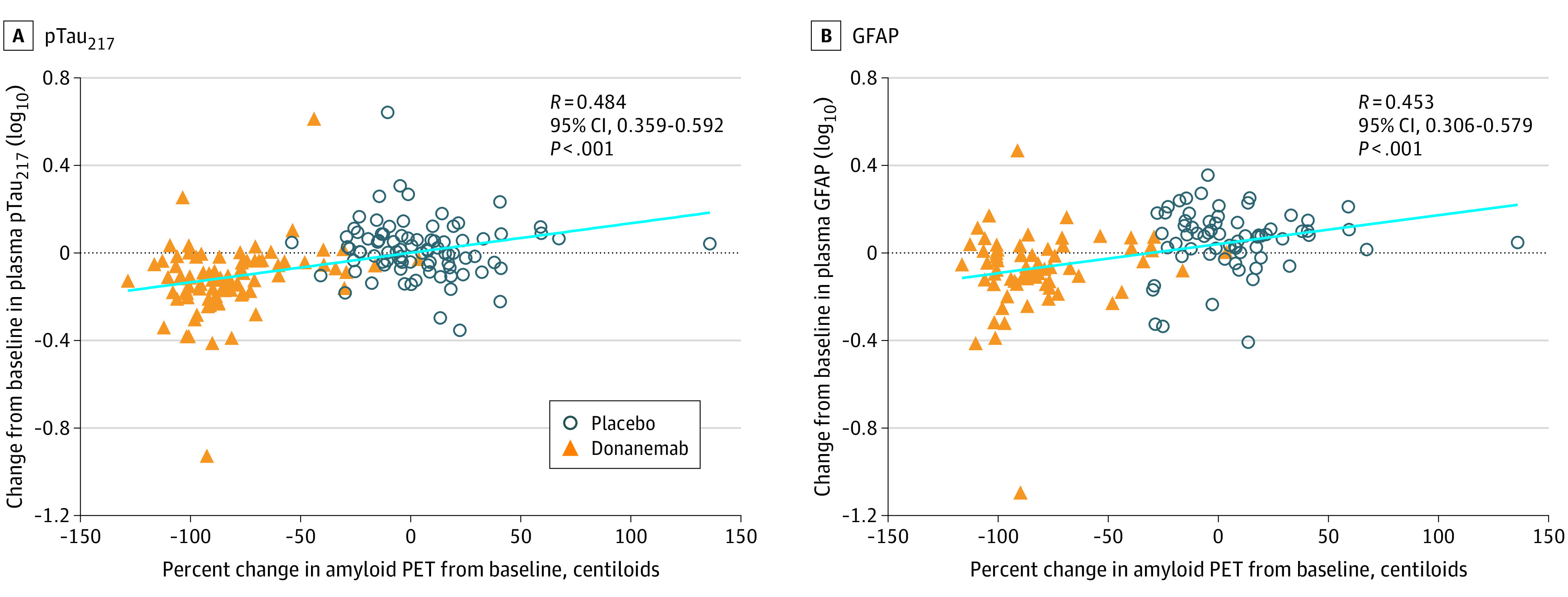
Figure 3. Correlations of Change in Plasma Phosphorylated tau217 (pTau217) and Glial Fibrillary Acidic Protein (GFAP) With Change in Amyloid Positron Emission Tomography (PET) End Points.

Percent change in amyloid PET levels from baseline to 76 weeks compared with change in plasma levels of pTau217 (placebo n = 85; donanemab n = 84) (A) and GFAP (placebo n = 66; donanemab n = 66) (B) from baseline to 76 weeks. Plasma levels were log10 transformed. Linear regression of all data points, regardless of treatment, is shown in light blue. Spearman rank was used for correlation coefficient.
