Figure 1.
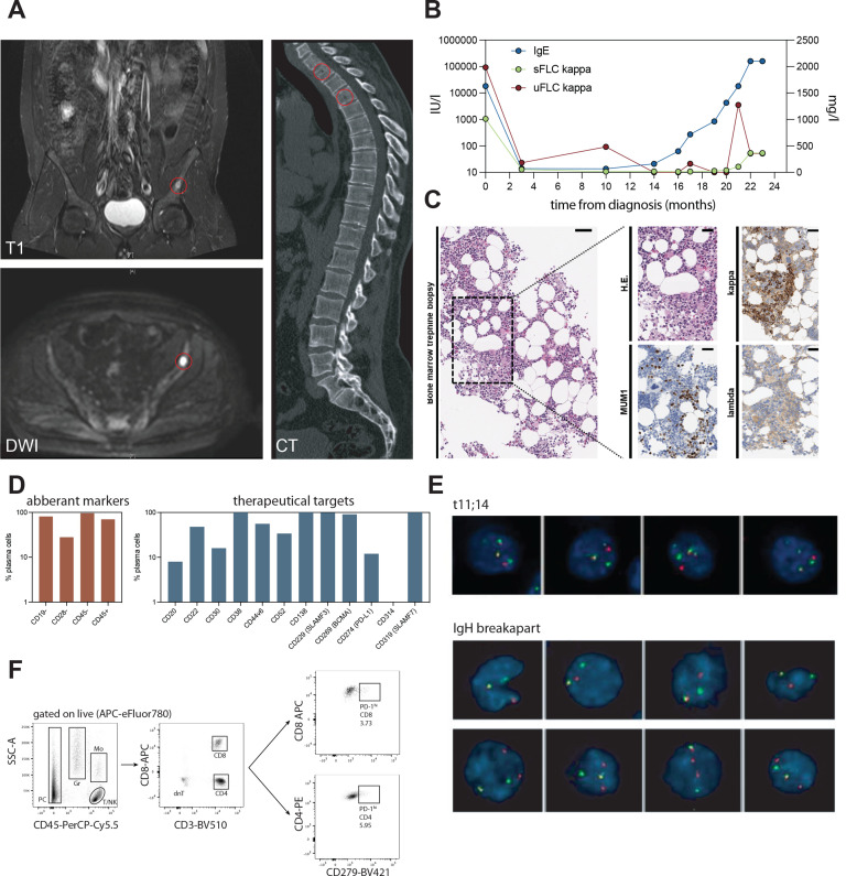
Clinical case presentation of IgE type multiple myeloma. (A) MRI (T1 and DWI) and CT imaging of osteolytic lesions in pelvic bone and cervical spine (highlighted in red). (B) Serological activity parameters (IgE in IU/L, serum free kappa light chains (sFLC) and urine free kappa light chains (uFLC) in mg/l) over course of disease. (C) Histopathological features of bone marrow biopsy at disease relapse. Immunohistochemistry stains indicated in-figure. (D) Flow cytometry quantification of aberrant markers and expression of potential therapeutic targets on malignant IgE type MM cells in bone marrow aspirate at relapse. (E) Cytogenetic analysis by iFISH identifies IgH rearrangement in form of t(11;14). (F) Flow cytometry-based assessment and quantification of bone marrow-associated CD4+ and CD8+ T cells and respective programmed cell death protein 1 (PD-1) surface expression on CD4+ and CD8+ T cell subsets. MM, multiple myeloma. DWI, diffusion weighted imaging.

