FIGURE 2.

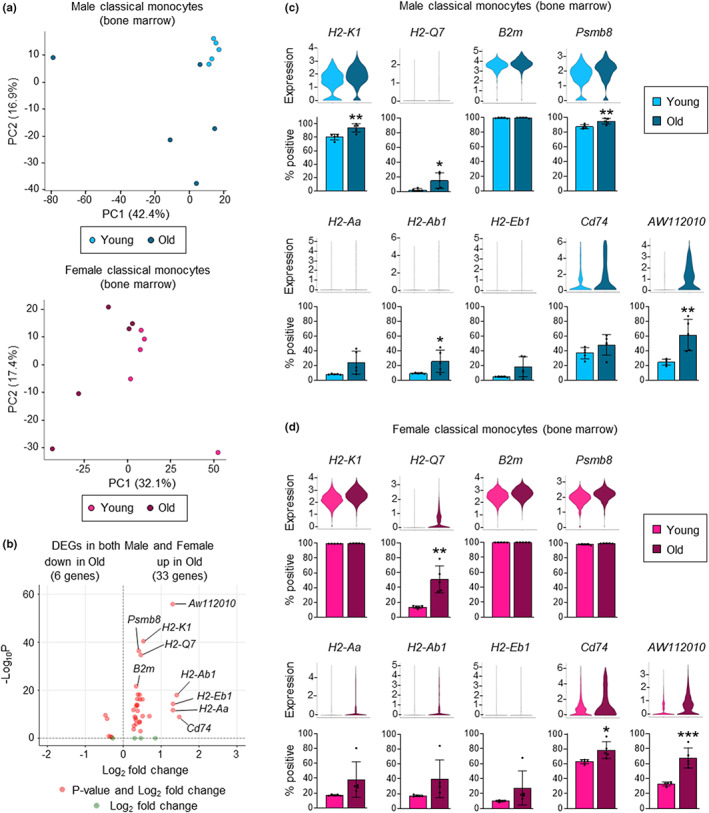
Aging increases the expression of genes associated with antigen presentation in bone marrow classical monocytes. scRNAseq analysis of classical (Ly6Chi) monocytes from the bone marrow of young and old, male and female mice (5 mice per group). (a) Principal component analysis of young and old, male (upper panel) and female (lower panel) mice. (b) Volcano plot of aging‐associated differentially expressed genes (DEGs; old vs. young) that are increased or decreased in both male and female mice. Aging‐associated DEGs were first defined separately using the male and female datasets (see Figure S4C–E) and then mean fold changes were calculated and Fisher's method was used to obtain combined adjusted p values. (c, d) Expression of DEGs (upper panels; all ‐Log10 P > 8) and percentage positive cells (lower panels) in young and old classical monocytes from male (c) and female (d) mice. Percentage positive cells are presented as mean plus standard deviation of 5 mice in each group, and statistical significance was assessed by two‐tailed Student's t‐test (*p < 0.05, **p < 0.01, ***p < 0.001, ****p < 0.0001).
