
FIGURE 6.
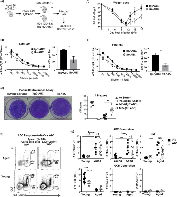
IgD+ABC produce neutralizing anti‐IAV Ab and inactivated IAV immunization generates iABC. (a) IgD+ABC (CD45.2) were sorted by flow cytometry and transferred into CD45.1 MD4 hosts and infected with 0.3 LD50 (25 PFU) PR8 (IAV). Hosts were sacrificed and serum was collected at 28–30 dpi (b) Kinetics (0–18 dpi) of weight loss between MD4 hosts with IgD+ABC and without IgD+ABC ELISA were performed with 28–30 dpi. Serum was collected from individual mice in both groups. (c) The titer of anti‐IAV‐specific IgM Ab was determined by ELISA. OD curves are shown over the titrated range (left) and the titer of anti‐IAV Ab (right) (d) The titer of anti‐IAV‐specific total IgG Ab OD curves is shown over the titrated range (left) and the titer of anti‐IAV Ab (right) (e) Plaque Neutralization Assay: using serum from the MD4 hosts with transferred IgD+ABC versus no ABC (n = 5–6 pooled from 2 to 3 separate experiments) Shown is an example of the plaque assay (left) and all the data from individual serum samples (right). (f) Experimental Design: Aged and young B6 mice were infected with 0.3 LD50 (25 PFU) PR8 (IAV) i.n. or treated with 5 μg whole inactivated PR8 (WIV) i.v. and sacrificed at 14 dpi. Representative FACS plots showing iABC and GCB responses in the spleen of aged and young B6 mice (g) The iABC (top) and GCB response (bottom) responses in spleen, lungs, and BM from individual mice in each group and is shown. Error bars represent SEM. Statistical significance determined by two‐tailed, unpaired Student's t‐test; *p < 0.05; **p < 0.01; ***p < 0.001, ****p < 0.0001.