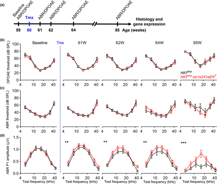
FIGURE 1.

Ntf3 overexpression at mid‐life slows the age‐related decline in ABR peak 1 amplitude but does not alter age‐related ABR and DPOAE threshold shifts. (a) Timeline of the experimental design. Ntf3 overexpressing (Ntf3 stop :Slc1a3/CreER T ) and control (Ntf3 stop ) mice were used in this study. Ntf3 overexpression was induced by intraperitoneal tamoxifen injection at 60 weeks of age (highlighted in blue). Baseline ABRs and DPOAEs were recorded 1 week before tamoxifen treatment (59 weeks of age) and at four later time points (61, 62, 64, and 95 weeks of age). After the last ABR and DPOAE tests, cochleae were collected and processed for either immunohistochemistry followed by confocal microscopy to evaluate HCs and IHC synapse counts or for RT‐qPCR to evaluate Ntf3 mRNA expression. (b) Ntf3 overexpression starting at 60 weeks of age does not alter DPOAE thresholds during the following 35 weeks. (c) Ntf3 overexpression starting at 60 weeks of age does not alter ABR thresholds during the following 35 weeks. (d) Ntf3 overexpression starting at 60 weeks of age increases ABR peak 1 amplitudes 1 week later and slows the decline of ABR peak 1 amplitudes at 80 dB SPL during the following 34 weeks. Ntf3 stop n = 7–13 mice; Ntf3 stop :Slc1a3/CreER T n = 7–17 mice. Two‐way ANOVA followed by Sidak's multiple comparisons test was used to evaluate statistical differences between Ntf3 stop and Ntf3 stop :Slc1a3/CreER T mice at every individual time point for either DPOAE threshold, ABR threshold or ABR peak1 amplitude. ** p < 0.01; *** p < 0.001. Error bars represent SEM
