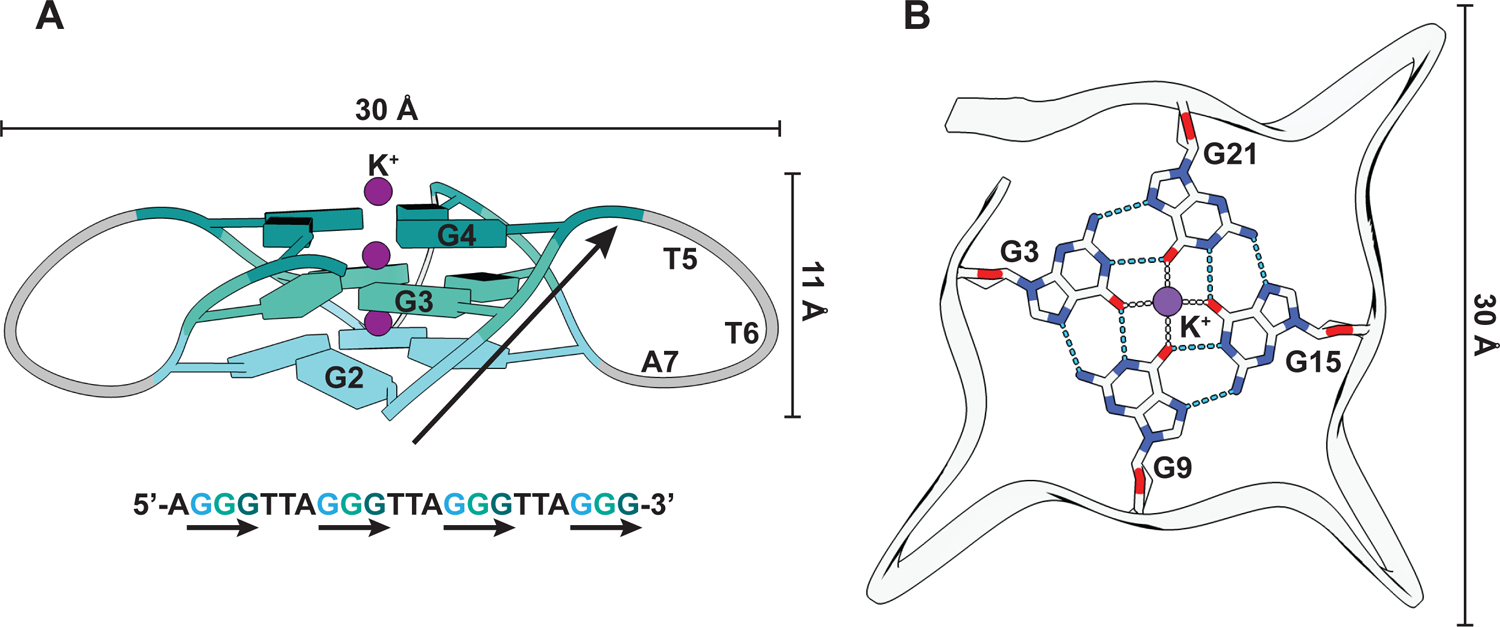
Figure 1: G-quadruplex structure.

(A) Structural representation of a G4 from human telomeric DNA (PDB: 1KF1) that folds into a parallel G4 structure (Parkinson, Lee, and Neidle 2002). The guanines are represented as slabs and colored differently per quartet. G-strands are marked with black arrows in both the structural model and the corresponding DNA sequence. (B) Interactions between the guanines of one quartet and with K+ ion (purple). Hydrogen bonds and ionic interactions are depicted as blue and gray dashed lines, respectively.
