FIGURE 10.
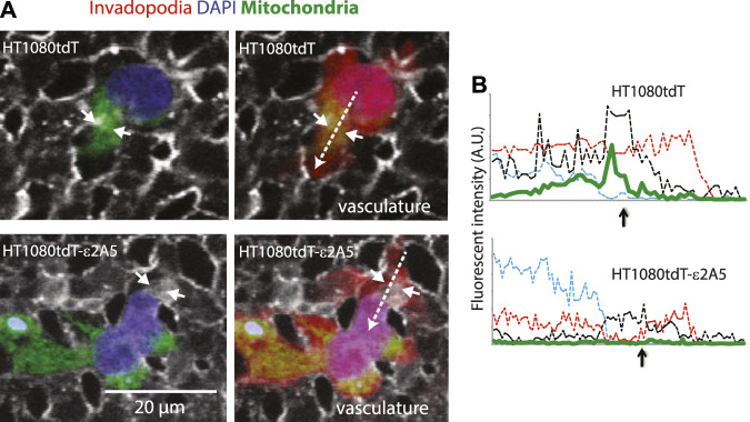
SLC2A5 function is necessary for polarized localization of mitochondria in HT1080tdT human fibrosarcoma cells in vitro and in vivo. (A) mitochondria (green) localization in HT1080tdT or HT1080tdT-ε2A5 (vasculature, grey; blue, DAPI) cells that are extravasating out of the chicken CAM vasculature (in vivo). Right panels show all three channels: red fluorescent protein (red), mitochondria (green), nuclei (blue) and vasculature (grey). Time-lapse video is shown in the Supplementary Video S3. (B). Fluorescent channel intensity along the line scans (dashed arrows in (A) (right panel) indicate the direction of the scan). Green line depicts mitochondria localization. The short arrows in (A) and (B) indicate vascular membrane breaches.

