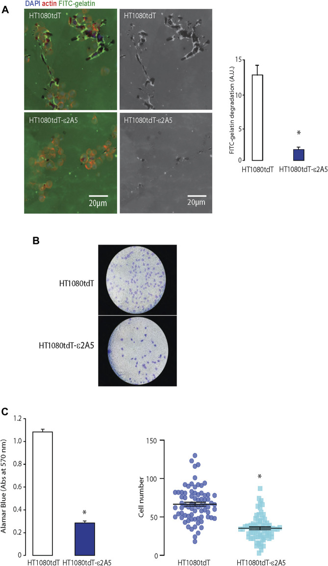
FIGURE 4.
Migration of the HT1080tdT-ε2A5 fibrosarcoma cells. (A). An FITC-gelatin degradation assay for invasiveness of HT1080tdT and HT1080tdT-ε2A5 cells. Samples were co-stained with DAPI and actin. *p < 0.0001, n = 3. (B). Transwell migration of HT1080tdT and HT1080tdT-ε2A5. Cells were fixed in 100% methanol and stained with Coomassie blue (n = 3). (C). Transmigration of HT1080tdT and HT1080tdT-ε2A5 across an 8 µm pore membrane. *p < 0.0001, n = 3. The images are representative of more than 3 biological replicates.

