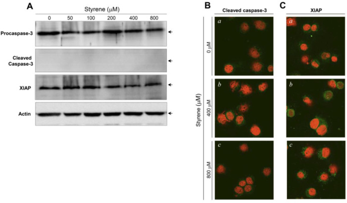
Fig. 3. Caspase-3 and Xiap proteins in TM3 Leydig cells after exposure to various concentrations of styrene.
(A) Western blot analysis of caspase-3, cleaved (activated) caspase-3 and Xiap after styrene treatment for 48 h. Equal amounts of protein (30 μg) were separated by SDS-PAGE and immunoblotted using a cleaved form specific antibody for corresponding proteins. Actin expression was examined as a loading control. Representative confocal images of the activated caspase-3 (B) and XIAP (C). Cells were treated with styrene for 48 h. The cells were then cytospun, fixed, and immunostained with an active form specific antibody for corresponding proteins. Green fluorescence (FITC), target proteins; Red fluorescence (PI), nucleus. a, Control; b, 400 μM styrene-treated; c, 800 μM styrene-treated. Original magnification: ×800.

