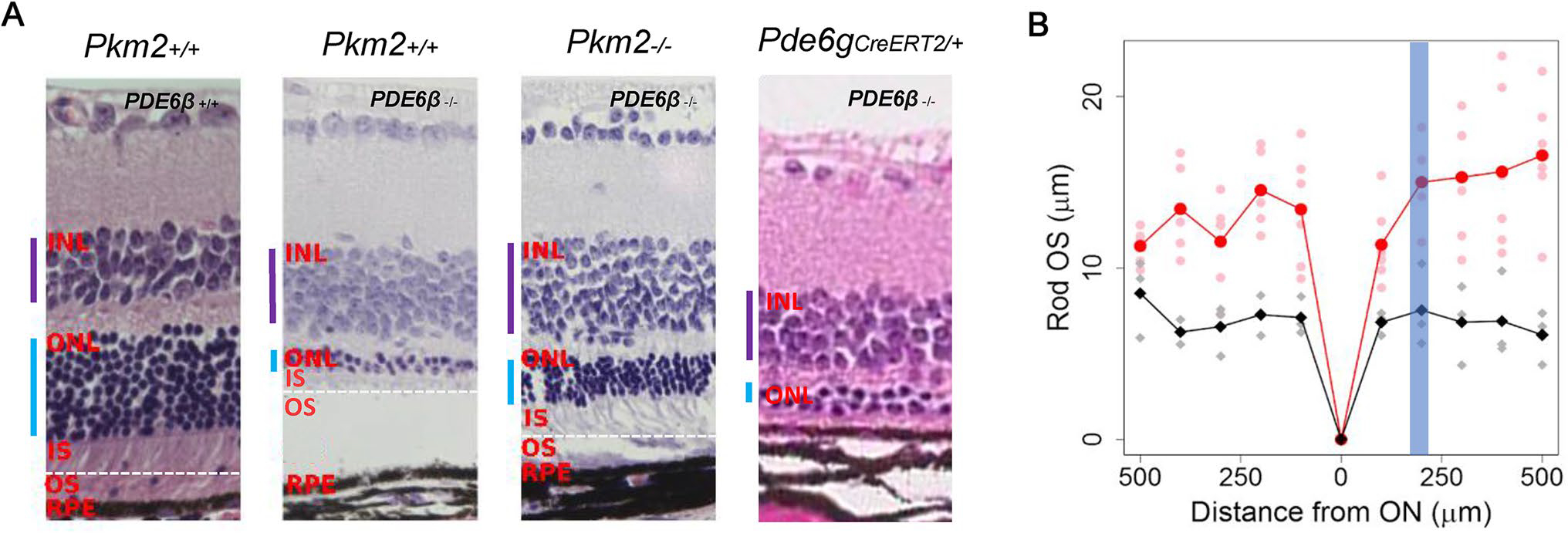
Fig. 2.

Deletion of Pkm2 in Pde6βH620Q/H620Q mice improved retinal morphology. a Representative H&E-stained retinal sections were imaged 180–220 μm from the optic nerve. Pde6gCreERT2/+ mice were imaged at P60, and all other mice were imaged at nine weeks (P64-P67). Selective deletion of Pkm2 enhanced but did not completely rescue ONL thickness in Pde6βH620Q/H620Q Pkm2−/− mice (n = 3), but not in Pkm2+/+ Pde6βH620Q/H620Q controls. At P8, P9, and P11, experimental and Pde6gCreERT2/+ mice were injected tamoxifen while wildtype and control mice were injected with oil. (Purple bar indicates INL, blue bar indicates ONL, dashed white horizontal line denotes IS/OS junction). b The difference in rod OS length was quantified at nine weeks (P64–P67). Selection deletion of PKM2 increases Rod OS thickness at every distance from the ON (n = 3). Red represents experimental mice, and black represents control mice. Highlighted line shows average Rod OS while faded dots represent individual measurements for mice. Blue band represents the area within which the histology images were taken (180–220 μm)
