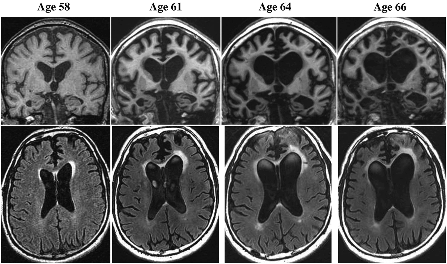
FIGURE 3–2.

Imaging of the patient in CASE 3-2. Representative coronal T1-weighted (top row) and axial fluid-attenuated inversion recovery (FLAIR) (bottom row) images at ages 58, 61, 64, and 66. The MRI at age 58 shows atrophy in the right amygdala and mesial frontal regions, along with mildly increased signal on FLAIR in the left more than right anterior periventricular regions, including the anterior corpus collosum. Over time, progressive atrophy in the frontal and temporal regions and associated ventricular dilatation are apparent, along with expansion of the increased signal particularly in the left anterior periventricular region.
