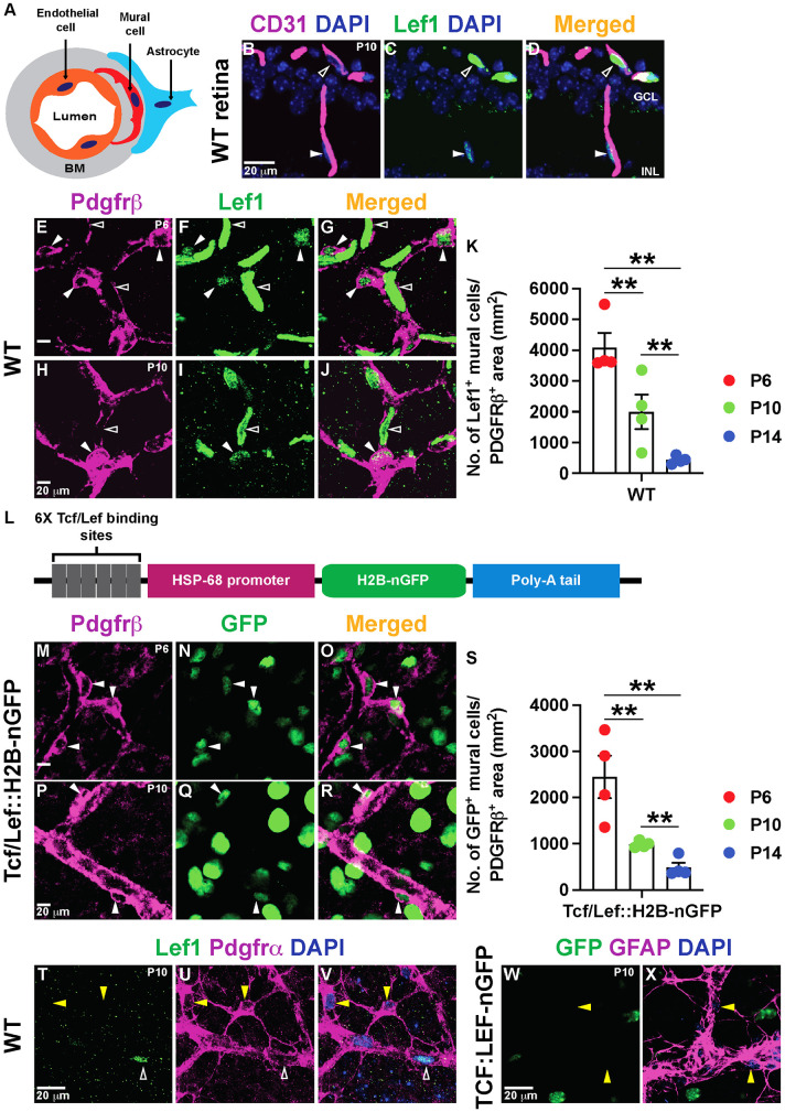
Fig. 1.
Retinal vascular mural cells have active Norrin/β-catenin signaling. (A) Schematic of the cell types in the NVU. The vBM is depicted in gray. (B-D) P10 WT retinal sections stained for Lef1 (green) and CD31 (magenta). Unfilled arrowheads indicate Lef1+ ECs and filled arrowheads indicate Lef1+ cells next to the ECs in the ganglion cell layer (GCL) and inner nuclear layer (INL). (E-J) Lef1+ ECs (unfilled arrowheads) and Lef1+ mural cells (filled arrowheads) in WT retinal flat-mounts stained for Lef1 (green) and Pdgfrβ (magenta). (K) Lef1+ mural cell numbers in the retina at three developmental time-points (n=4/mice per time point). (L) Schematic of the Tcf/Lef::H2B-nGFP transgene. (M-R) Tcf/Lef::H2B-nGFP retinal flat-mounts labeled for GFP (green) and Pdgfrβ (magenta). Filled arrowheads indicate GFP+ mural cells. (S) GFP+ mural cell numbers in the retina at the indicated developmental time-points (n=4/mice per time point). (T-V) P10 WT retinal flat-mounts stained for Lef1, Pdgfrα and DAPI. Yellow arrowheads indicate Lef1− astrocytes. Unfilled arrowheads indicate Lef1+ ECs. (W,X) P10 Tcf/Lef::H2B-nGFP retinal flat-mounts stained for GFP, GFAP and DAPI. Yellow arrowheads indicate GFP− astrocytes. Data are mean±s.e.m., analyzed with one-way ANOVA with Bonferroni corrections: **P<0.02.

