Fig. 2.
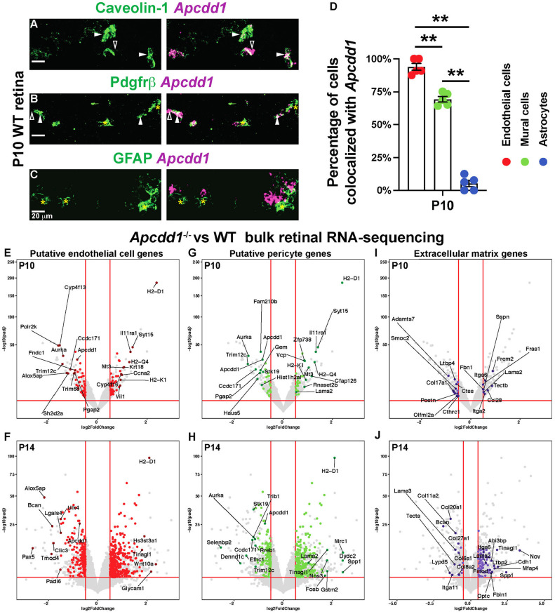
Apcdd1 is expressed in both retinal ECs and PCs and Lama2 expression is increased in Apcdd1−/− retinas. (A-C) FISH of P10 WT retinal sections with antisense mRNA probes against Apcdd1 (magenta) followed by immunostaining for caveolin 1, Pdgfrβ or GFAP (green). Filled arrowheads, asterisks and unfilled arrowheads indicate Apcdd1 inside the cell, Apcdd1− cells and Apcdd1 outside the cell, respectively. (D) Fraction of Apcdd1+ ECs, mural cells and astrocytes in the retina (n=4 mice). (E-J) Volcano plots of putative EC (E,F: red dots), PC (G,H: green dots) and ECM (I,J; purple dots) genes differentially expressed between P10 and P14 Apcdd1−/− and WT retinas. Genes above the horizontal red lines are those with significantly different expression in Apcdd1−/− retinas. Genes to the right of vertical red lines are significantly upregulated, whereas those to the left are significantly downregulated in Apcdd1−/− retinas. The most differentially expressed genes are named. Data are mean±s.e.m., analyzed with two-tailed unpaired Student's t-test: **P<0.02.

