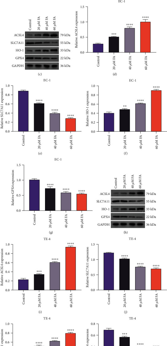
Figure 5.

FA exposure contributes to ferroptotic cell death of ESCC cells. (a, b) Iron content was detected in EC-1 and TE-4 cells with 48 h administration of 20 μM, 40 μM, and 60 μM FA through iron content kit. (c–g) Activities of ACSL4, SLC7A11, HO-1, and GPX4 were measured in EC-1 cells following exposure to 20 μM, 40 μM, and 60 μM FA for 48 h through immunoblotting. (h–l) Activities of ACSL4, SLC7A11, HO-1, and GPX4 were tested in TE-4 cells that were administrated with 20 μM, 40 μM, and 60 μM FA for 48 h via adopting immunoblotting. p was computed through one-way ANOVA test. Significance level was denoted as ∗p < 0.05, ∗∗p < 0.01, ∗∗∗p < 0.001, and ∗∗∗∗p < 0.0001.
