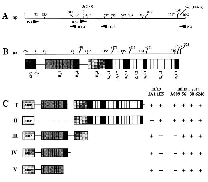
FIG. 1.
Schematic representation of the VspA fusion proteins and localization of immunodominant epitopes 1A1 and 1E5. (A) Localization of the primers on the vspA gene used to generate the recombinant proteins represented in panel C (numbers indicate nucleotide position). (B) Representation of the different domains that compose the VspA product (open boxes) and their localization (numbers represent amino acid position). (C) Representation of the domains that compose the FP-VspA-I to -V fusion proteins and their reactivity with MAbs 1E5 and 1A1 and animal sera A009 (experiment 2), 56 and 30 (from two animals of outbreak 4), and 6248 (from one animal of outbreak 2).

