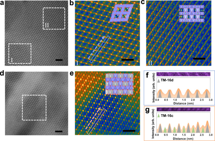
Fig. 3. Ex situ STEM investigations of Li1.46Ni0.32Mn1.2O4–x-based electrodes. a–c.
HADDF-STEM images of the as-synthesized CD-LNMO powder (a), and FFT-filtered images of the I (b), and II (c) regions. d, e HADDF-STEM images of the fully charged CD-LNMO powder. e FFT-filtered images of the marked region in d. f, g Enlarged HADDF-STEM images and corresponding signal profiles of the regions marked with blue and orange boxes in b and e, respectively. The occupancies of TM ions in the 16d and 16c sites are represented by gray and green triangles, respectively. Scale bars in a, d and b, c, e represent 2 and 1 nm, respectively.

