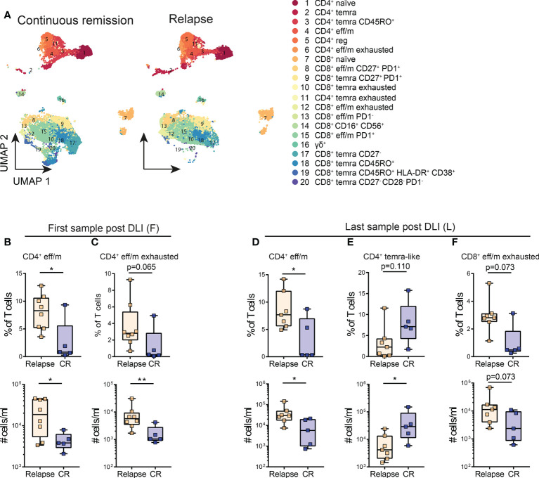
Figure 3.
Lower levels of exhausted T cell clusters are associated with continuous remission early and late post DLI. (A) Cluster analysis identified 20 different T cell populations within the post-DLI samples. These are shown in Uniform Manifold Approximation and Projection (UMAP) including all samples of patients with continuous remission (n=5) and relapse (n=8) post DLI during the 24 months follow-up. The details on the 20 identified clusters are listed in Supplementary Table S6 . (B, C) Comparison of frequencies and cell counts of two clusters identified in patient samples early after DLI between patients with continuous remission (blue, n=5) and relapse (orange, n=8) during the 24 months study follow-up. (B) Comparison of CD4+ eff/m and (C) exhausted CD4+ eff/m clusters between the groups. (D–F) Comparison of frequencies and cell counts of three clusters identified in patient samples late after DLI between patients with continuous remission (blue, n=5) and relapse (orange, n=7) during the 24 months study follow-up. (D) Comparison of CD4+ eff/m, (E) CD4+ temra CD45RO+ and (F) exhausted CD8+ eff/m clusters between the groups. All Clusters are named as in (A). (B–F) Lines represent median, boxes represent 25th and 75th percentile and whiskers represent minimum and maximum. Statistical analysis (B–F), was performed by Student t test or Mann-Whitney test (twotailed). *p<0.05, **p<0.001.

