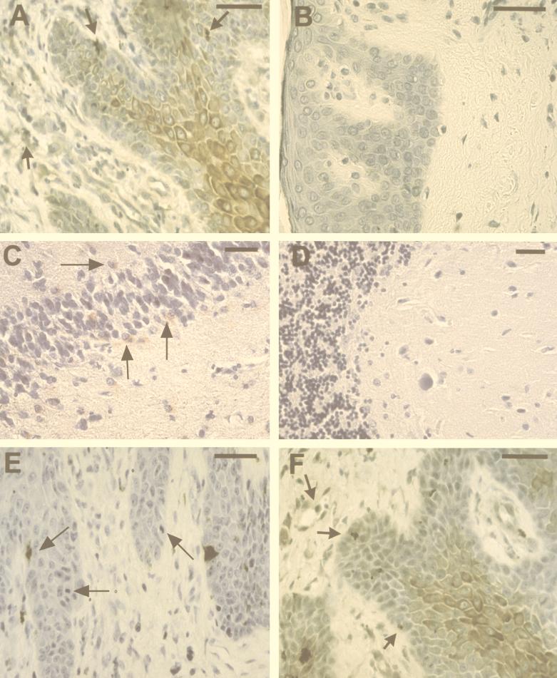
FIG. 2.
Immunohistochemical analysis of anti-SAF, anti-C. pneumoniae OMP, and anti-Chlamydia LPS reactivity in formalin-fixed skin and brain tissue. Anti-SAF immunoreactivity is shown in panels A to D. (A) Immunolabeling of epidermal cells with the morphology of keratinocytes, as well as inclusion-like structures and perivascular cells (arrows). (B) A section from a mycosis fungoides patient found negative for immunolabeling (negative control). (C) Anti-SAF (67 μg/ml) reactivity on temporal cortex of a patient with AD confirmed positive for C. pneumoniae in other studies (positive control) (4). Immunolabeling of apparent glial cells in the denate gyrus is depicted by arrows. (D) Negative immunoreactivity of anti-SAF is shown on a section from the cerebellum (uninvolved) of the same patient (negative control). (E) Immunolabeling of inclusion-like structures with anti-OMP MAb (1:10) (arrows) of a section from a patient with mycosis fungoides. (F) Immunoreactivity of anti-LPS (1:10) of cells with the morphology of keratinocytes as well as labeling of inclusion-like structures and perivascular cells (arrows). Diaminobenzidine-cobalt (dark brown) chromagen was used for panels A, B, E, and F. 3-Amino-9-ethylcarbazole (red) chromagen was used for panels C and D. Bars = 50 μm.

