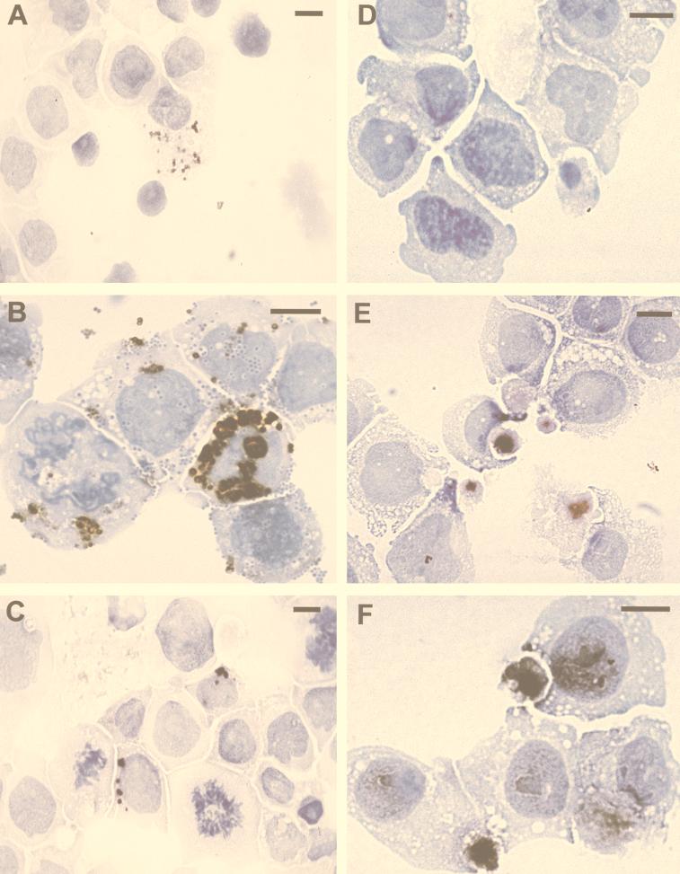
FIG. 3.
Immunolabeling of THP-1 monocyte cell line infected with C. pneumoniae isolated from a brain from a patient with AD and with the laboratory strain of C. pneumoniae (TW-183). Cultured THP-1 cells were placed on slides by a cytospin and fixed with STF. THP-1 cells infected with C. pneumoniae isolated from the brain of a patient with AD are shown in panels A to C: immunolabeling of anti-OMP (A), anti-SAF (B), and anti-LPS (C) of bacterial inclusions within infected cells. (D) Uninfected THP-1 cells that reacted with anti-SAF (negative control). THP-1 cells infected for 10 days with the laboratory strain of C. pneumoniae TW-183 immunolabeled with anti-OMP (E) or anti-SAF (F) contain diffusely stained inclusions. Bars = 10 μm.

