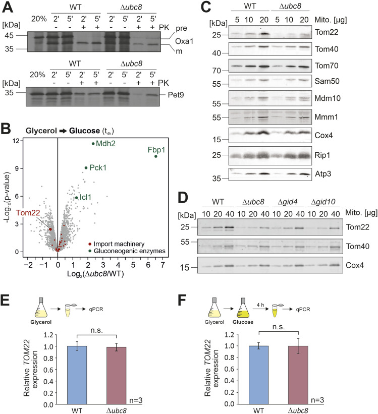
Figure 4. Absence of Ubc8 or other components of the GID complex leads to diminished levels of the outer membrane protein Tom22.
(A) Radiolabeled Oxa1 and Pet9 were incubated with mitochondria isolated from wild-type or Δubc8 cells grown in galactose medium at 30°C for different times. Non-imported proteins were degraded by the addition of proteinase K (PK). 20% of the radiolabeled proteins used per import reaction are loaded for control. Proteins were visualized by autoradiography. (B) Cells were grown in a glycerol medium before glucose was added for 4 h. Cells were subjected to mass spectrometry, and the levels of different groups of mitochondrial proteins were analyzed. Four replicates were analyzed. See also Fig S5A and B and Table S3. (C, D) Cells were grown in a glycerol medium. After the addition of glucose for 4 h, mitochondria were isolated and subjected to SDS–PAGE. The indicated proteins were visualized by Western blotting. (E, F) TOM22 mRNA levels of cells continuously grown in glycerol (E) or after the addition of glucose for 4 h (F) were analyzed by qRT-PCR. Shown is the TOM22 expression in Δubc8 cells relative to the expression in wild-type cells. Mean values and standard deviations of three biological replicates (n = 3) were calculated. Significance testing was performed with a t test (n.s., not significant).
Source data are available for this figure.

