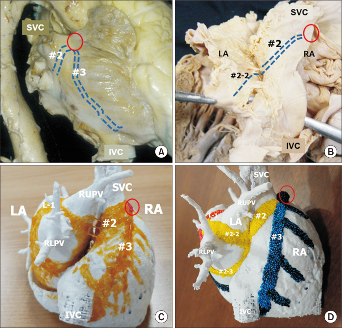
Fig. 2.
The major inter-nodal routes at the right lateral aspect of the atrial wall. These routes are broad bands or sheets, as shown in (C) and (D), although they are shown as lines in (A) and (B) to demonstrate the shape of the atrial wall. (A) Right lateral view of the heart showing external indicators of the middle and posterior inter-nodal routes from the sinus node (red circle). The middle route is a broad myocardial bundle at the sinus venarum. (B) After dissection of the Waterstone groove, the inter-atrial muscular connection is noted at the superior limbus of the oval fossa. The main stem of the middle inter-nodal route gives rise to the atrioventricular node at the superior limbus (#2-1) and then to the branches at the left atrial wall (#2-2,3). (C) The atrial chambers from the posterior aspect show the middle (second) inter-nodal route (#2) and the posterior (third) route at the crista terminalis (#3). (D) The middle inter-nodal route is colored yellow, and the posterior inert-nodal tract is colored blue. The middle route branches to form sub-branches extending to the left atrial wall (#2-2, #2-3). The posterior inter-nodal route gives rise to the pectinate muscles and approaches the atrioventricular node and the coronary sinus. Fig. 2C is modified from the authors’ previous publication [25]. SVC, superior caval vein; IVC, inferior caval vein; LA, left atrium; RA, right atrium; RLPV, right lower pulmonary vein.

