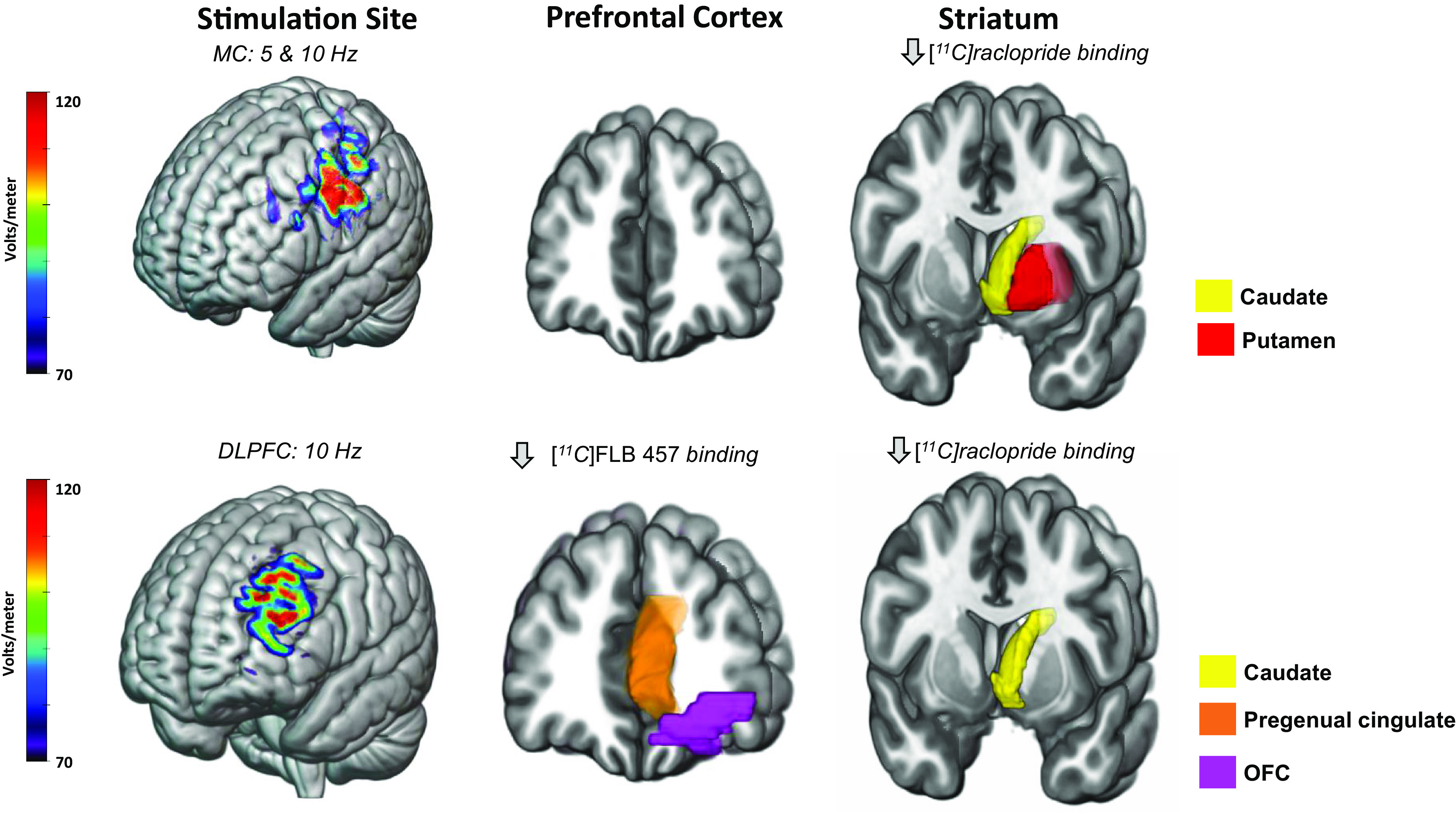
Fig. 3.

TMS to the motor cortex and the DLPFC influences dopamine receptor availability in a region-specific manner. Above are representative models of electric fields following TMS to the motor (top) and dorsolateral prefrontal (bottom) cortices in standard space. D2 dopamine receptor availability was measured in vivo using [11C]raclopride, a radiotracer detectible through PET scanning procedures. Reported above are regions exhibiting a decrease in [11C]raclopride binding in at least two publications. One study used [11C]FLB 457. Overall, 5 Hz and 10 Hz TMS to the left motor cortex decreases [11C]raclopride binding in the caudate (yellow) and putamen (red). 10 Hz TMS to the left dorsolateral prefrontal cortex (DLPFC) decreases [11C]FLB 457 binding in the pregenual cingulate (orange) and orbitofrontal cortex (OFC) (purple). Additionally, 10 Hz TMS to the left DLPFC decreases [11C]raclopride binding in the caudate (yellow) and putamen (red). Decreases in dopamine receptor availability suggest a TMS-induced dopamine release. Modeling parameters include a Magstim B70 coil at 60% machine output and standard tissue conductivity values. The electric fields depicted range from 70 to 120 millivolts.
