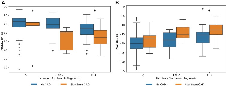
Figure 3.
Box plots of artificial intelligence quantification of (A) left ventricular ejection fraction and (B) global longitudinal strain at peak stress stratified by ischaemic burden and presence of coronary artery disease. * P < 0.001 for ≥3 ischaemic segments; the number of cases for 0 and 1–2 ischaemic segments was too low for statistically assessing for significant differences.

