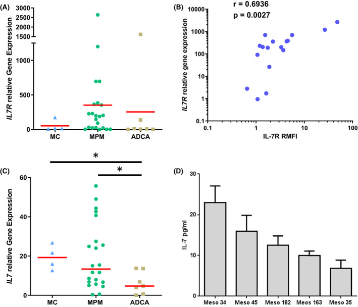
Fig. 1.

Analysis of IL‐7R and IL‐7 expression in MPM cells. (A, C) mRNA expression of IL7R (A) and IL7 (C) measured by RT‐PCR in MPM (n = 22), ADCA (n = 7), and primary mesothelial cells (n = 4). (B) Correlation between IL7R mRNA expression and IL‐7Rα cell surface expression measured by flow cytometry in MPM cells (n = 17). Spearman test. (D) IL‐7 secretion by MPM cells as measured by ELISA of cell culture supernatants. Red horizontal bars represent median expression. Bar graphs represent mean ± SEM of three independent experiments. (A, C) Mann–Whitney t‐test. *P < 0.05. ADCA, lung adenocarcinoma; MC, mesothelial cell; MPM, malignant pleural mesothelioma.
