
Fig. 8.
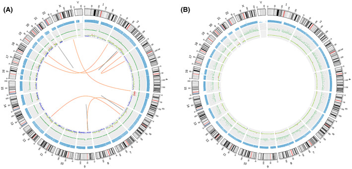
Genomic variation circos display in the primary lymphoepithelioma‐like carcinoma of the renal pelvis. (A) Genomic variation circus for case 1. (B) Genomic variation circus for case 2. The five‐layer structure from the outside to the inside represents the sequencing coverage map, the density of karyotype stripe, single‐nucleotide variant, insertions and deletion, copy number variation, and the structural variation results, respectively. All the experiments were repeated thrice independently.