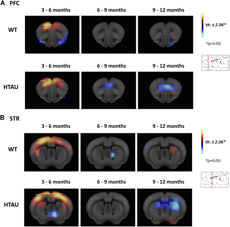
FIGURE 2.
Age-related changes in brain metabolism in htau mice. In vivo brain glucose uptake was analysed by [18F]-FDG preclinical PET imaging in htau and WT mice at 3-, 6-, 9- and 12-month-old. The result is shown as a statistical parametric comparison between the groups. Color changes represent a statistical change, determined by t-test comparisons (*p < 0.05) between the groups compared (indicated in each panel). Red-yellow look up table side indicates [18F]-FDG uptake increases, while blue indicates uptake decrease. Coronal sections showing [18F]-FDG uptake changes in the Prefrontal cortex (A) and striatum (B) of htau and WT mice, between 3 and 6, 6 to 9 and 6–12 months old (WT n = 11; htau n = 9). Images shown represent T-maps of brain metabolism compared to former recorded activity of the same group.

