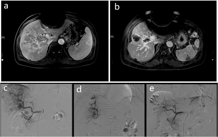
Figure 1.
These MR and DSA images show the imaging data of a 68-year-old men with HCC complicated by portal vein tumor thrombosis in the right branch. Contrast-enhanced MR scan showing a tumor thrombus in right portal vein (A). The contrast-enhanced MR of the patient 3 months later shows a significant reduction in PVTT volume (B), and the previously blocked portal vein due to PVTT also restores blood flow. At the same time, according to the mRECIST criteria, there was no activity in the intrahepatic lesions, and no activity was found in the PVTT, which was judged as CR. The DSA image of the patient undergoing embolization of the APFs and obvious portal vein development can be seen during angiography of the proper hepatic artery (C). The angiographic image of the patient taken at a time after treatment, showing that the APFs are completed by sealing (D). A follow-up DSA image of the patient three months later showed no APFs (E).

