Figure 3.
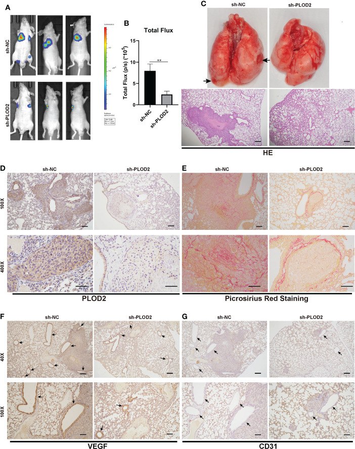
PLOD2 promotes OS metastasis and angiogenesis in vivo. (A, B). 143B cells transfected with a lentiviral vector sh-PLOD2 or sh-NC were injected into nude mice via the tail vein (1 × 10⁵ cells per mice, n = 3 each group). Representative image and analysis of luminescence intensity in tail vein-injected mouse models. (C). The lung specimen and HE staining of metastatic lung nodules. The HE staining samples were imaged at 40× magnification. Scale bar = 250 μm. (D). IHC of metastatic lung nodules in sh-NC and sh-PLOD2 group, the expression of PLOD2 was analyzed based on IHC results. The samples were imaged at 100× magnification, Scale bar = 100 μm and 400× magnification, Scale bar = 50 μm. (E). Picrosirius Red Staining of metastatic tumor in the lung. The samples were imaged at 100× magnification, Scale bar = 100 μm and 400× magnification, Scale bar = 50 μm. (F, G). IHC of metastatic tumors in the lung. The small blood vessels were marked with black arrows. The expression of angiogenesis protein VEGF and CD31 was analyzed. The samples were imaged at 40× magnification, Scale bar = 250 μm and 100× magnification, Scale bar = 100 μm. All data are presented as the means ± SD, **P < 0.01. (student’s t test).

