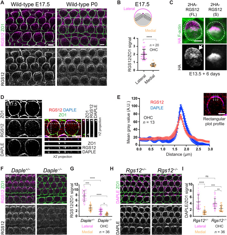
Fig. 6. RGS12 and DAPLE colocalize at the apicolateral HC junction.
(A) RGS12 and ZO1 coimmunolabeling. (B) RGS12 signal as a ratio of ZO1 at the lateral and medial HC junction. Means ± SD. n = HCs, N = 3 animals. Mann-Whitney test, ****P < 0.0001. (C) HA labeling of representative OHCs expressing the full-length (FL; Rgs12-201) or short (S; Rgs12-204) Rgs12 isoform (see fig. S6, C and D). The cochlea was electroporated at E13.5 and cultured for 6 days. 2HA-Rgs12 (FL) but not (S) is enriched at the lateral HC junction (arrow). Outcome representative of >18 OHCs in >4 explants. (D and E) RGS12 and DAPLE coimmunolabeling along with ZO1 as junctional marker in OHCs. In (D), side projections in the YZ (right) and XZ (bottom) axis are shown along with surface views. RGS12 and DAPLE signals are polarized and colocalize at the lateral HC junction. (E) Plot profile of averaged signal intensity along lines crossing the junction as illustrated. Means ± SEM. N = 3 animals. (F and G) RGS12 immunolabeling. RGS12 signals are reduced and less markedly polarized laterally in Daple mutants (F), as quantified in (G) for OHCs. (H and I) DAPLE immunolabeling. DAPLE junctional amounts are normal but less polarized in Rgs12 mutant OHCs (H), as quantified in (I). (G and I) Means ± SD. N = 3 animals. Kruskal-Wallis test with Dunn’s multiple comparisons. (G) ****P < 0.0001, ***P = 0.0001, and *P = 0.0266. (I) ns, P > 0.9999; ****P < 0.0001; ***P = 0.0004; and **P = 0.0021. Scale bars, 5 μm.

