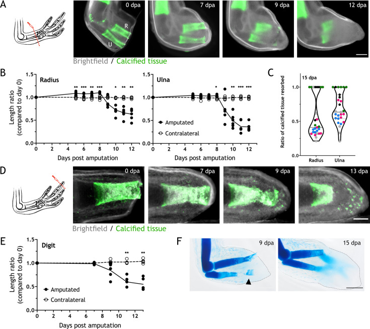
Figure 1. Skeletal elements are resorbed upon amputation.
(A) Time course of resorption during zeugopod regeneration. Calcein-stained axolotls were amputated at the distal end of the calcified tissue. R: radius; U: ulna; Scale bar: 500µm. (B) Quantification of resorption rate in radius and ulna in (A). Length ratio was calculated using the length at 0 dpa as a reference. Each dot represents an animal (n=6; *** p<0.001, ** p<0.01, * p<0.05, two-way ANOVA, Bonferroni’s multiple comparisons test, amputated versus contralateral). (C) Quantification of resorption percentage in calcified radius and ulna among animals in different assays. Each assay is represented by a color (pool of five independent experiments, n=27). (D) Time course of resorption during digit regeneration. Calcein-stained axolotls were amputated at the distal end of the calcified tissue. Scale bar: 200µm. (E) Quantification of calcified digit resorption in (D). Length ratio was calculated using the length at 0 dpa as a reference. Each dot represents an animal (n=5; ** p<0.01, two-way ANOVA, Bonferroni’s multiple comparisons test, amputated versus contralateral). (F) Alcian blue staining of limbs at different dpa. Arrowhead: broken piece of ulna. Dashed line: outline of distal limb. Scale bar: 500µm (n=2).

