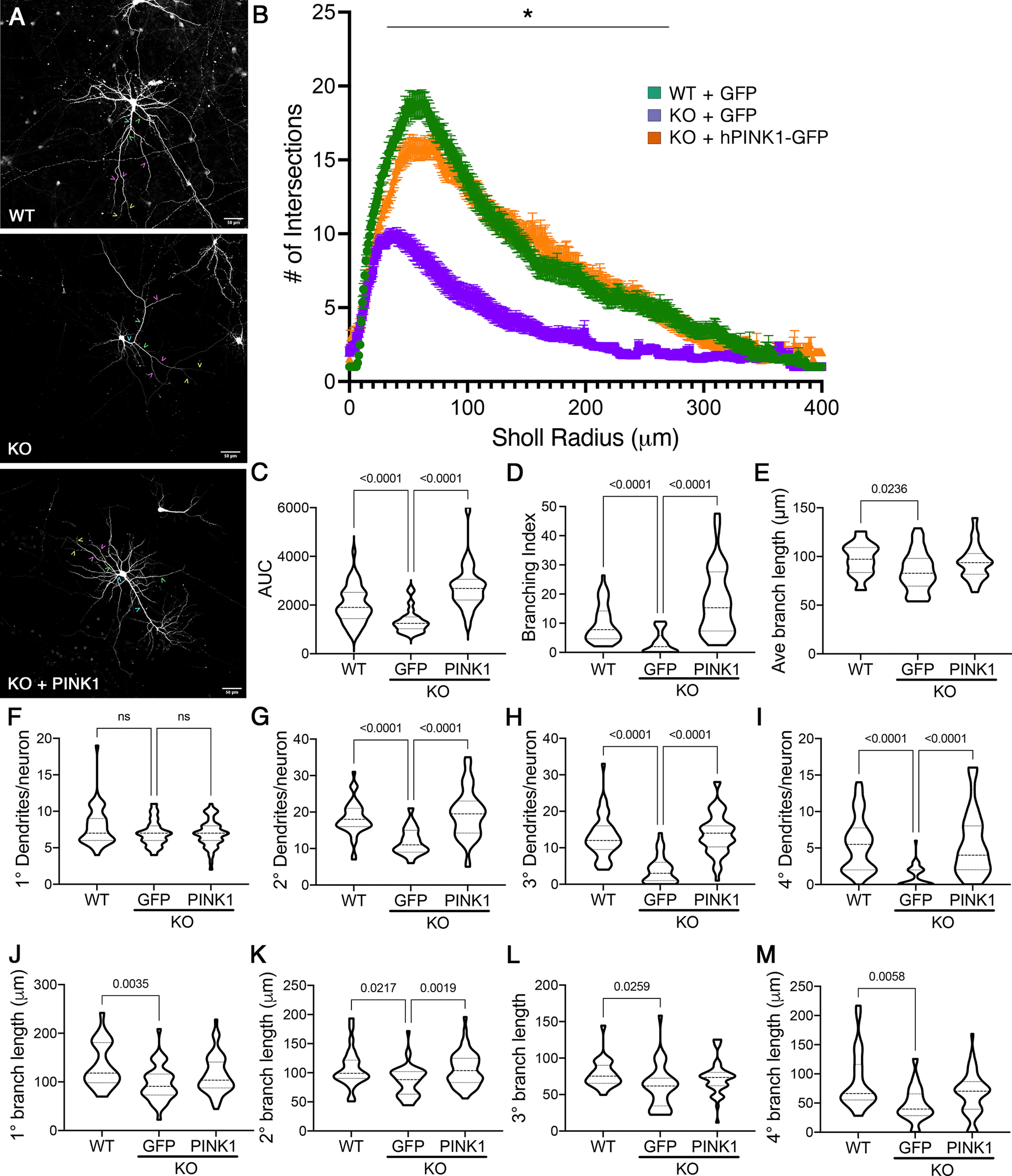
Figure 1.

Loss of endogenous PINK1 elicits dendritic simplification in primary cortical neurons, which is reversed by introduction of human PINK1. A, Primary cortical neurons from Pink1−/− (KO) mice were transfected with GFP or GFP+PINK1-GFP at low efficiency to enable visualization of their arbors and compared with WT controls transfected with GFP. Scale bar, 50 µm. Representative examples of 1° dendrites are marked with teal carets, 2° with green carets, 3° with pink carets, and 4° with yellow carets. B, Sholl analysis of dendritic arbors expressed as mean ± SEM; *radii with significant differences for KO versus WT and KO versus KO plus hPINK1 (multiple-comparison testing following two-way repeated-measures ANOVA; Table 2). C–E, AUC, branching index, and average branch lengths, respectively. F–I, The number of primary, secondary, tertiary, and quaternary branches per neuron. J–M, The average branch lengths of primary, secondary, tertiary, and quaternary dendrites per neuron. Data are expressed as violin plot probability densities with the median and interquartile range indicated. Adjusted p values following post hoc Dunnett's T3 multiple comparisons test are shown (n = 25–52 neurons per condition compiled from 3–4 independent experiments; Table 2). ns - not significant.
