Figure 1.

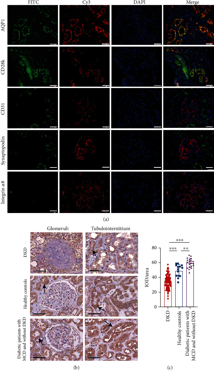
The expression of GPX4 in renal specimens. (a) GPX4 (marked with FITC-conjugated secondary antibody) mainly expressed in proximal and distal tubular epithelial cells (marked as AQP1 and CD28k with Cy3-conjugated secondary antibody, respectively), and there was little, if any, expression of GPX4 in glomerular podocytes and endothelial and mesangial cells (marked as synaptopodin, CD31, and integrin α8 with Cy3-conjugated secondary antibody, respectively). Renal specimens came from healthy controls. Scale bar = 50 μm. (b, c) Representative photographs (b) and semiquantification (c) of GPX4 IHC staining in DKD patients (n = 85), MCD patients with T2DM (n = 20), and healthy controls (n = 14). The black arrow indicates the staining of GPX4, and the scale bar = 50 μm (b). Horizontal lines represent mean ± SD (c). AQP1: Aquaporin 1; CD28k: Calbindin D28k; DKD: diabetic kidney disease; IOD/area: the staining intensity of GPX4; MCD: minimal change disease. ∗p < 0.05, ∗∗p < 0.01, and ∗∗∗p < 0.001.
