Fig. 1.
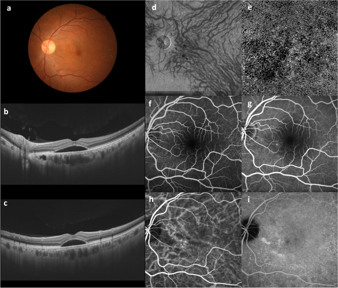
Characteristics of pachychoroid diseases. An illustrative case: 53-year-old man with central serous chorioretinopathy. a Color fundus photograph shows a serous retinal detachment (SRD) in the macula. b and c Horizontal and vertical 12-mm B-mode ocular coherence tomography (OCT) images through the fovea show pachychoroid with dilated outer choroidal vessels (vortex veins) associated with the SRD. The central choroidal thickness is 361 mm. d En face OCT image (12 × 12 mm) shows dilated vortex veins in the deep layer of the choroid. The horizontal watershed is lost due to the anastomoses between the superior and inferior vortex veins. e OCT angiography (3 × 3 mm) shows a decreased flow signal of choriocapillaris in the macula. f and g Fluorescein angiography (early and late phases) shows dye leakage within the SRD area. h and i Indocyanine green angiography (early and late phases) shows dilated choroidal vessels with hyperpermeability (pachyvessels) between the papilla and macula. Reproduced with permission from reference 31

