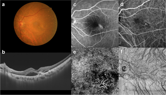
Fig. 11.
An illustrative case: images of the right eye of an 84-year-old female with polypoidal choroidal vasculopathy. a The color fundus photograph shows retinal pigment epithelium (RPE) detachments associated with subretinal hemorrhage and serous retinal detachment (SRD) in the macular area. b The 12-mm horizontal B-mode optical coherence tomography (OCT) images through the fovea show dilated outer choroidal vessels (vortex veins) accompanied by RPE detachment and SRD. The central choroidal thickness is 281 µm. c Fluorescein angiography shows window defects and leakage in the macular area. d Indocyanine green angiography shows dilated choroidal vessels and a polypoidal lesion temporal to the fovea. e OCT angiography (3 mm × 3 mm) shows network vessels accompanied by a polypoidal lesion between the detached RPE and Bruch’s membrane. f En face OCT image shows dilated vortex veins in the deep layer of the choroid. The horizontal watershed has been lost due to anastomoses between the superior and inferior vortex veins. Reproduced with permission from reference 33

