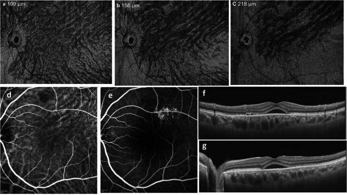
Fig. 5.
An asymmetric dilated vortex vein in central serous chorioretinopathy is shown. The left eye of a 48-year-old man with + 1.0 diopter of hyperopia. a En face OCT image in the superficial Haller’s layer at 109 μm from the Bruch membrane shows asymmetric superior vortex vein dilatation. b In the middle of the Haller’s layer at 156 μm from the Bruch membrane, asymmetric dilatation of the superior vortex vein is more clearly pronounced. Vortex vein branches do not taper toward the posterior pole. The horizontal watershed zone cannot be seen. c Deeper in the Haller’s layer, at 218 μm from the Bruch membrane. The superior vortex veins are dilated and retain their large calibers, terminating abruptly at their distal ends. d The venous phase of indocyanine green angiography (ICGA) shows hyperpermeability corresponding to the dilated superior vortex vein. e Fluorescein angiography shows hyperfluorescence of the retinal pigment epithelium in the area of hyperpermeability in ICGA images. f A vertical B-mode scan shows an enlarged vascular lumen of the choroid in the upper half of the posterior pole. g A horizontal B-mode scan shows dilated vortex veins at the horizontal watershed zone. Reproduced with permission from reference 19

