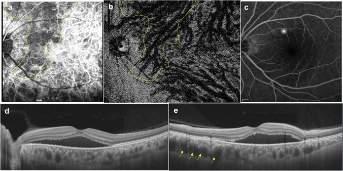
Fig. 6.
A 39-year-old man with acute central serous chorioretinopathy. a Early-phase indocyanine green angiography (ICGA) shows a geographic area (yellow line) corresponding to a filling delay in the choriocapillaris. b En face OCT image of the Haller’s layer of the choroid shows an asymmetrically dilated superior vortex vein the location of which (yellow line) corresponds to the geographic filling delay on ICGA. c Fluorescein angiography shows dye leakage from the region of a dilated vortex vein. d A horizontal B-mode scan shows serous retinal detachment with pachychoroid (central choroidal thickness; 459 μm). e A vertical B-mode scan shows marked dilatation of the superior vortex vein (arrows). Reproduced with permission from reference 21

