Fig. 7.
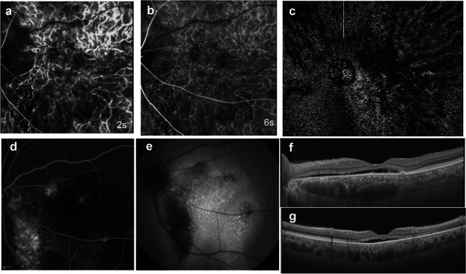
A 48-year-old man with chronic central serous chorioretinopathy. a Early-phase indocyanine green angiography (ICGA) shows a geographic area of filling delay in the choriocapillaris. b The choroidal filling delay persists in the venous phase. c En face OCT image shows anastomosis between superior and inferior vortex veins in the macular area. Anastomotic vessels and surrounding vortex veins show dilatation. The region of these dilated vessels corresponds to the area of the filling delay on ICGA. d Late-phase fluorescein angiography shows leakage above the macula and hyperfluorescence in the descending tract. e Fundus autofluorescence shows hyperfluorescent dots in the area of serous retinal detachment and hypofluorescence in the descending tract. f A horizontal OCT B-mode scan shows serous retinal detachment with dilated vortex and anastomotic veins between the papilla and the macula. g A vertical B-mode scan shows dilated vortex veins. Reproduced with permission from reference 21

