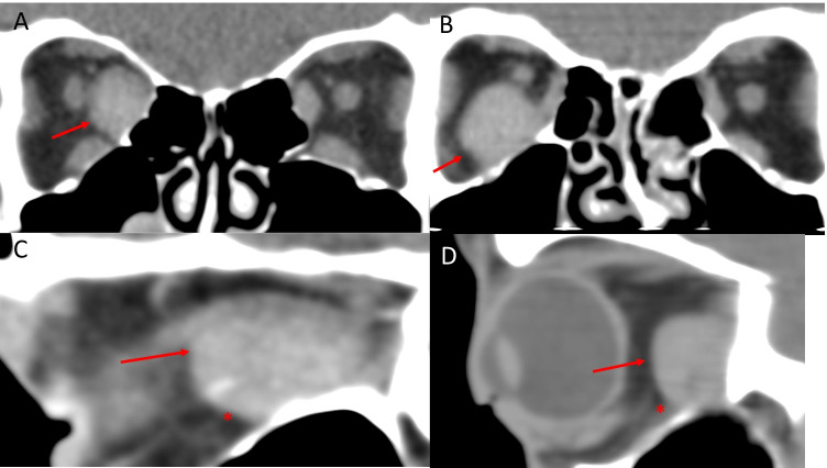
Fig. 15.
Two cases of Spontaneous haemorrhage related to extraocular muscles. Case 1—coronal (A) and sagittal (C) CT scans showing a large, well-defined mass in the right medial rectus with an anterior rounded border (arrow) and posterior tapering edge (*) towards the orbital apex. The sagittal image has a ‘bleached whale’ appearance suggestive of intramuscular haemorrhage. Case 2—coronal (B) and sagittal (D) CT scans showing a large, well-defined mass in the right inferior rectus with an anterior rounded border (arrow) and posterior tapering edge (*) towards the orbital apex

